Phaeohyphomycosis refers to a group of mycoses (fungal infections) that are dematiaceous, which means they are pigmented. The pigment is due to their ability to deposit melanin in their cell walls.
Histology of phaeohyphomycosis
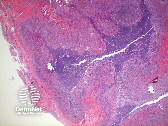
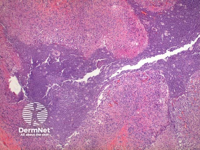
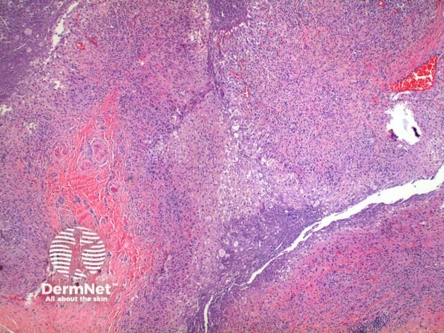
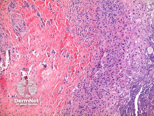
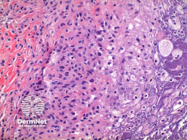
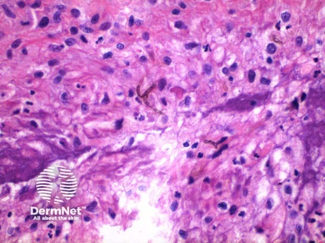
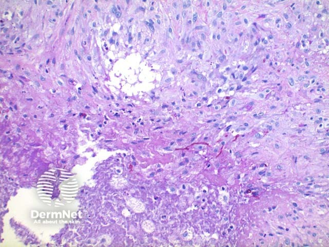
Scanning power view of phaeohyphomycosis shows a deeply extending granulomatous pattern (Figure 1) which may show areas of necrosis (Figure 2). Centrally an abscess or cysticnodule may form. Frequently a foreign body such as a wood splinter can be seen. The epidermis commonly shows pseudoepitheliomatoushyperplasia. The inflammatoryinfiltrate is comprised of histiocytes with multinucleatedgiant cells, and numerous neutrophils (Figures 3,4 and 5). At high power branching septate pigmented fungal hyphae can be seen (Figure 6).
Phaeohyphomycosis pathology
Figure 1
Figure 2
Figure 3
Figure 4
Figure 5
Figure 6
Special stains in phaeohyphomycosis
PAS (Figures 7 and 8) or Grocott methenamine silver stains can be used to enhance visualisation of the fungal hyphae.
Phaeohyphomycosis pathology, PAS stain
Figure 7
Figure 8
Differential diagnosis of phaeohyphomycosis
Chromomycosis: This pigmented fungal infection demonstrates formation of rounded sclerotic or septate bodies/muriform cells.
References
Skin Pathology (3rd edition, 2002). Weedon D
Pathology of the Skin (3rd edition, 2005). McKee PH, J. Calonje JE, Granter SR