In dermoscopy, perpendicular white lines are short discrete white lines oriented parallel and orthogonal (perpendicular) to each other and seen only under polarised light [1]. They are also known as polarising white lines, short white lines, shiny white lines, shiny white streaks, chrysalis, chrysalids, and crystalline structures. Perpendicular white lines are a clue to a specific diagnosis including basal cell carcinoma (BCC) and some melanomas [2].
What do perpendicular white lines look like through the dermatoscope?
Perpendicular white lines are only seen under polarised light. They appear as short, shiny white lines and move as the dermoscopy lens is moved at different angles over the lesion.
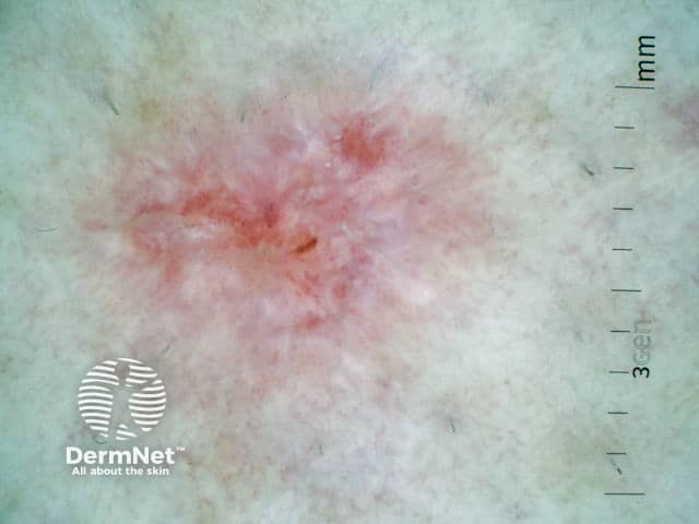
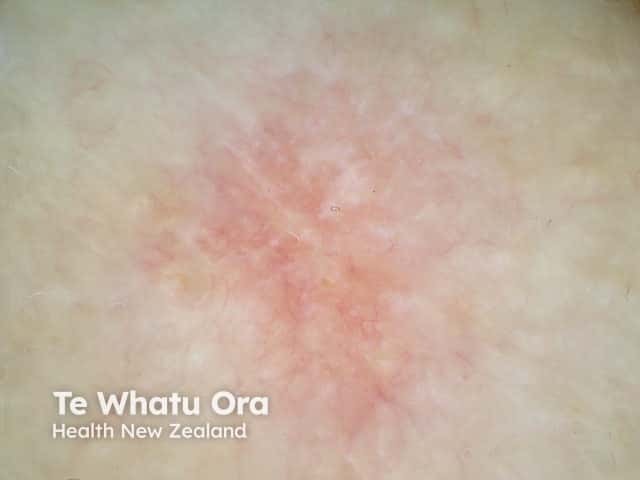
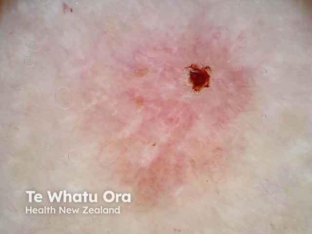
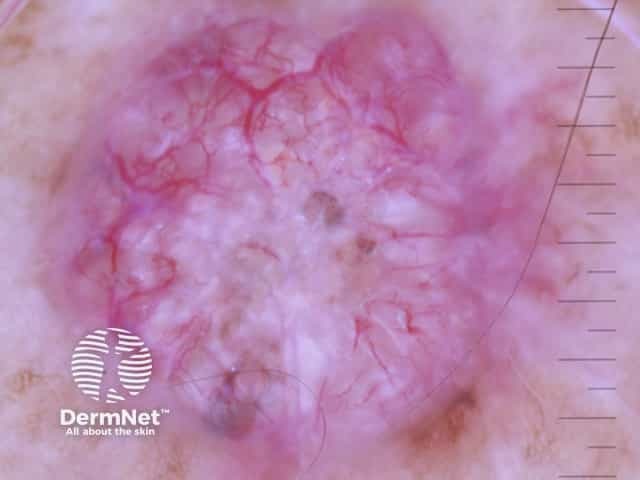
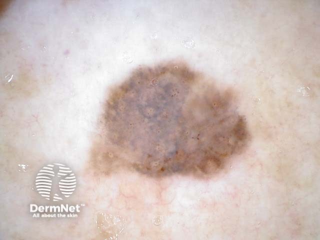
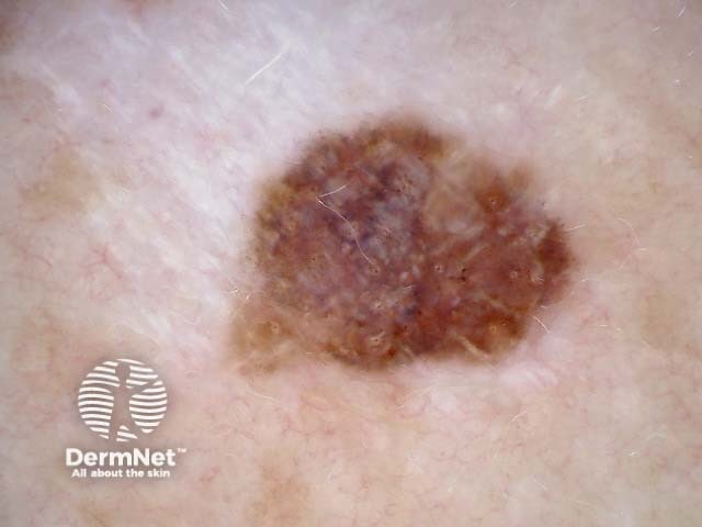
Perpendicular white lines in BCCs
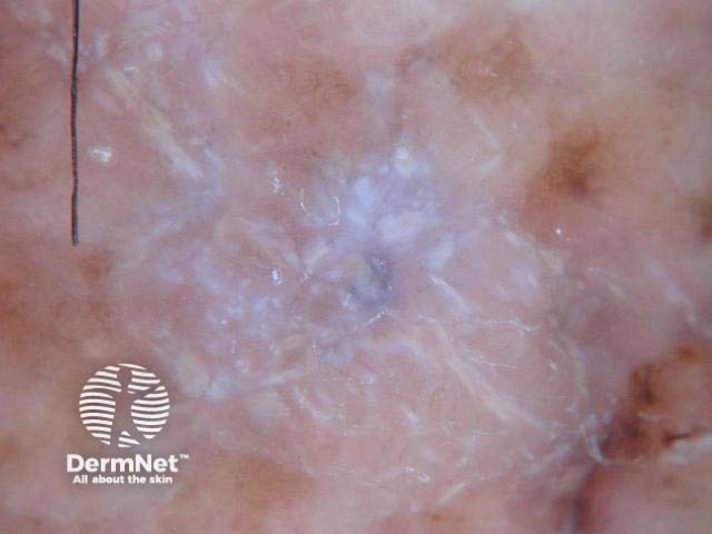
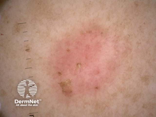
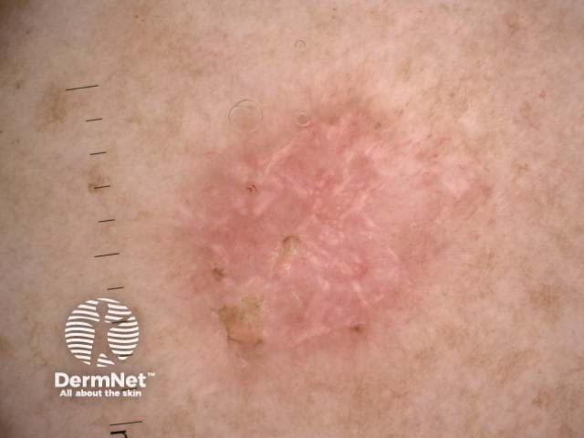
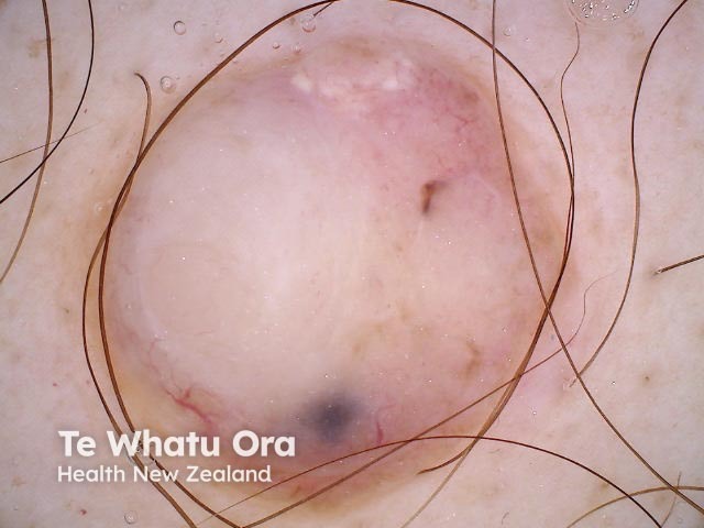
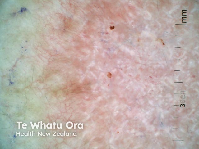
Superficial basal cell carcinoma dermoscopy
Superficial basal cell carcinoma dermoscopy
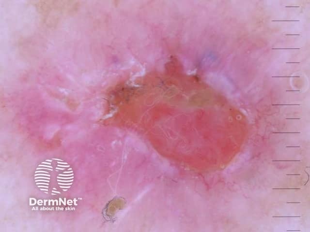
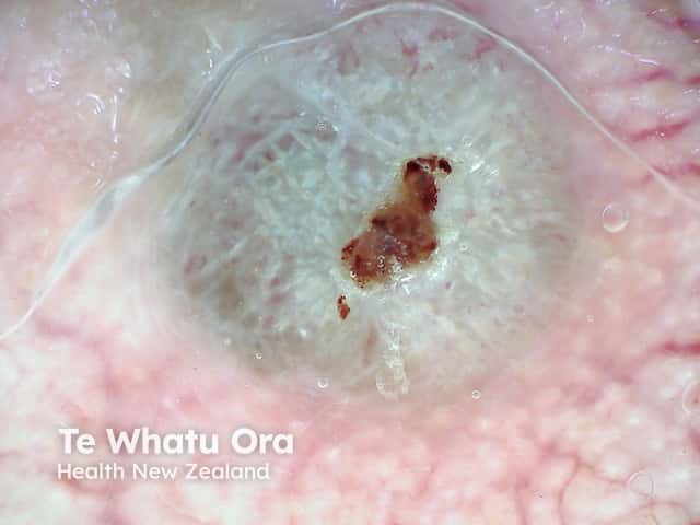
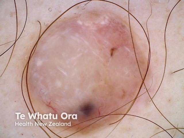
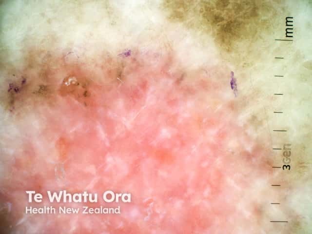
Nodular basal cell carcinoma dermoscopy
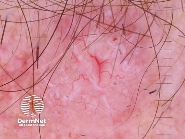
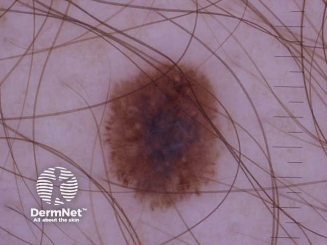
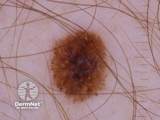
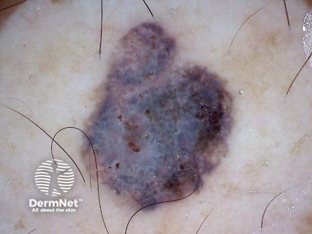
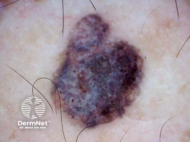
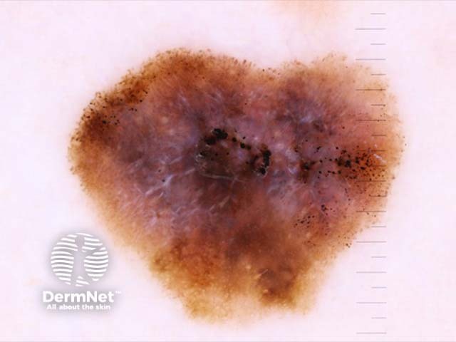
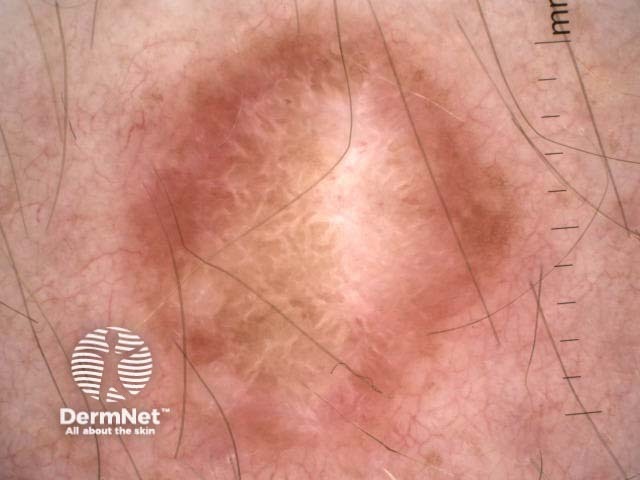
Perpendicular white lines in melanoma
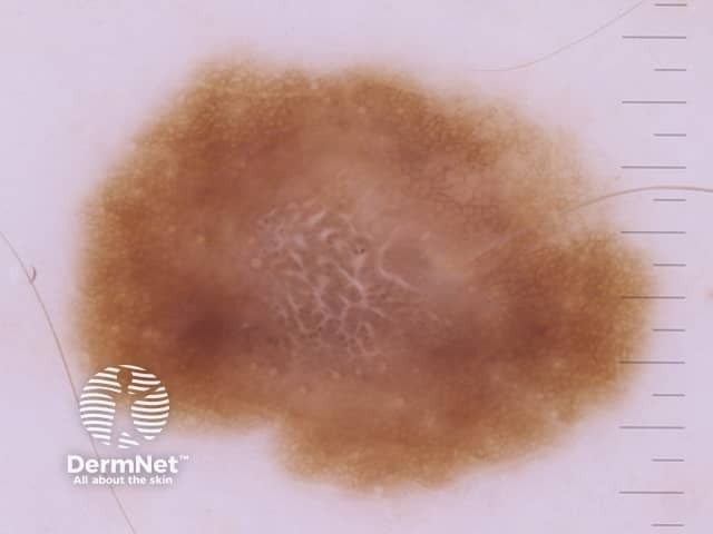
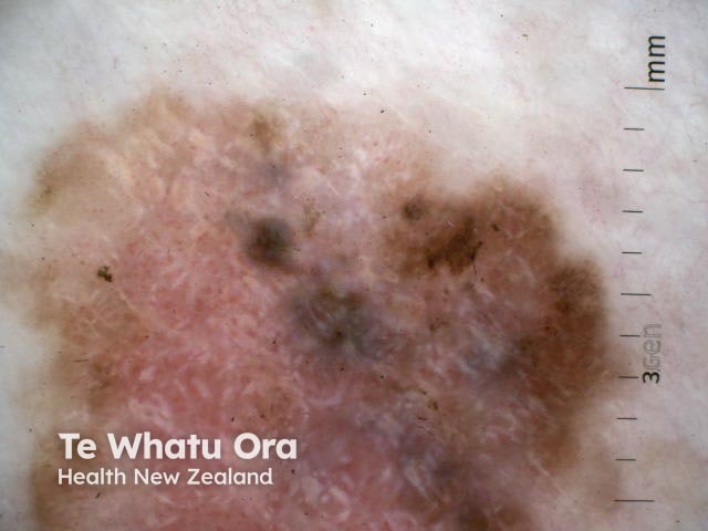
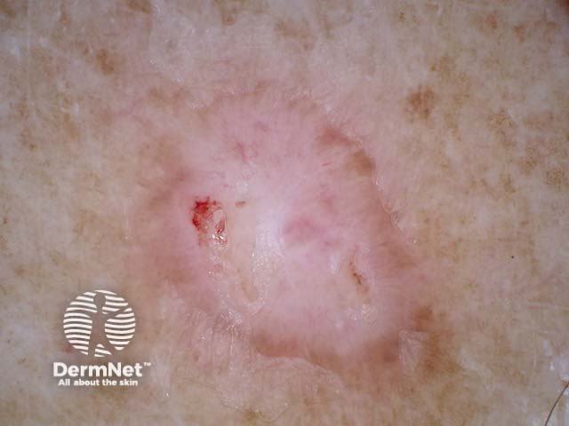
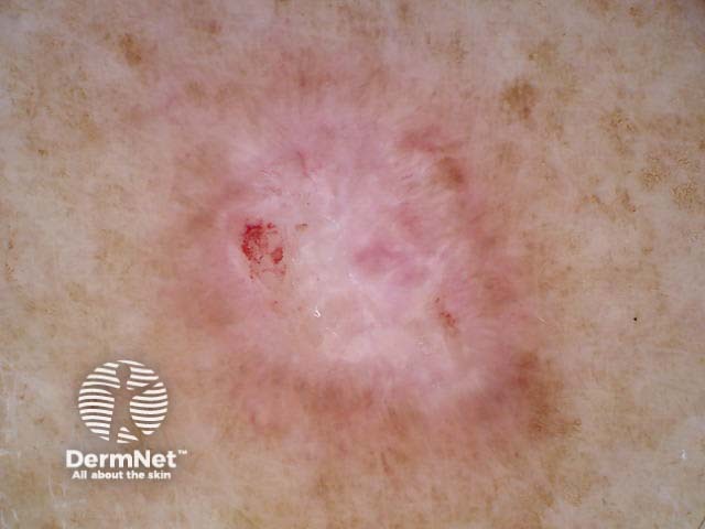
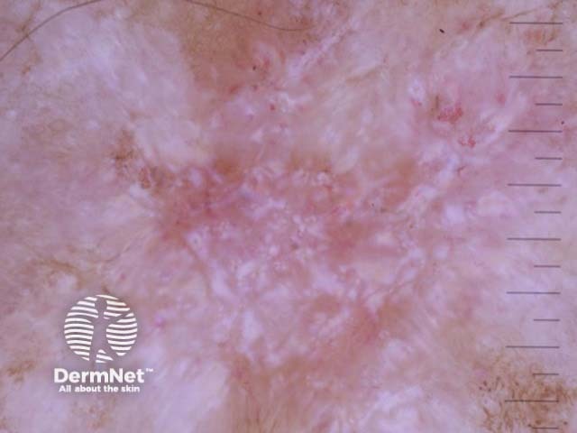
Melanomain situ dermoscopy
Melanoma in situ dermoscopy
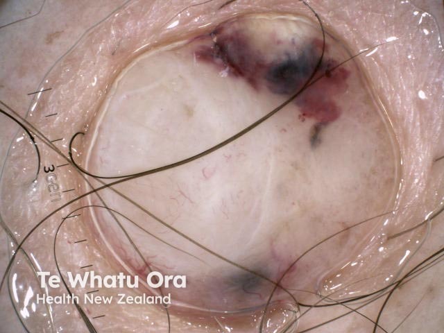
A nodular melanoma arising within a superficial spreading melanoma
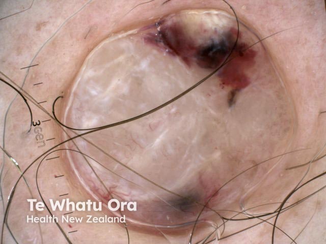
Amelanotic nodular melanoma
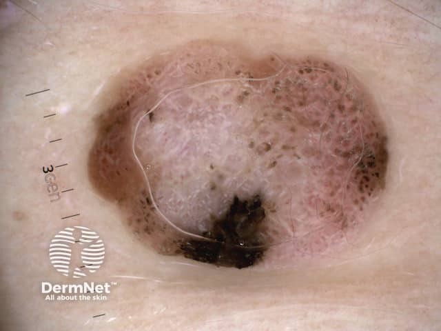
Nodular melanoma, Breslow 6.8 mm
Nodular melanoma, Breslow 2.5 mm, polarised dermoscopy view
Polarised and non-polarised light
The following pairs of images demonstrate the differences seen in dermoscopy of perpendicular white lines under polarised and nonpolarised light.
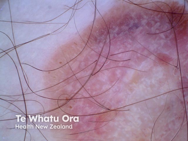
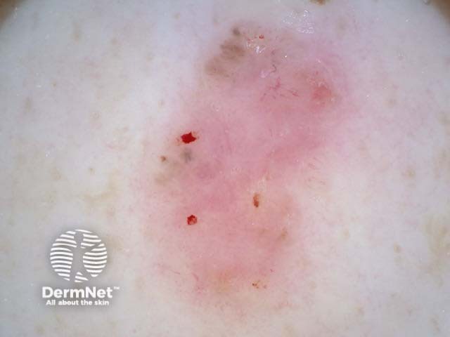
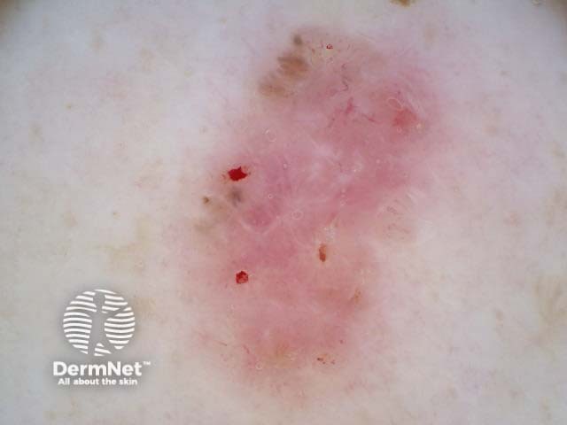
Polarised and nonpolarised light in BCC
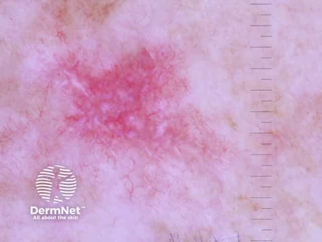
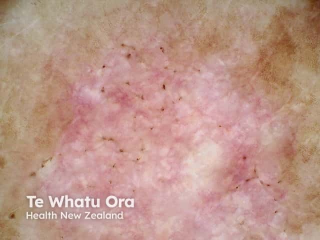
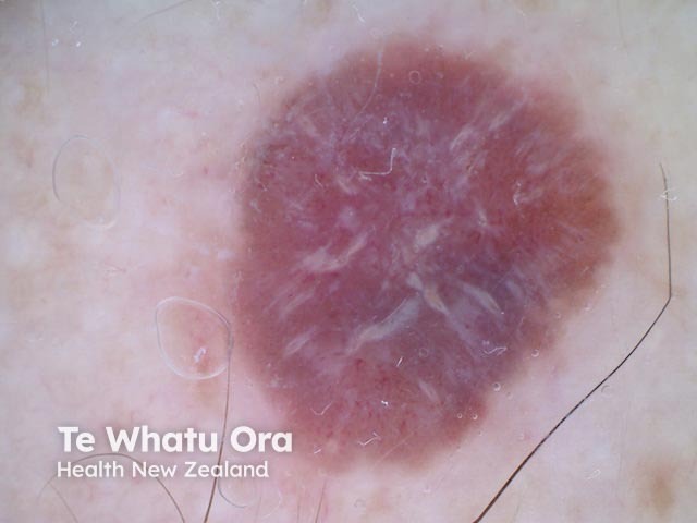
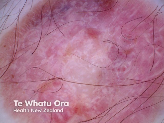
Polarised and nonpolarised light in melanoma
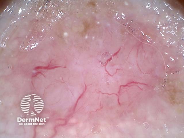
Melanoma in situ, nonpolarised dermoscopy view
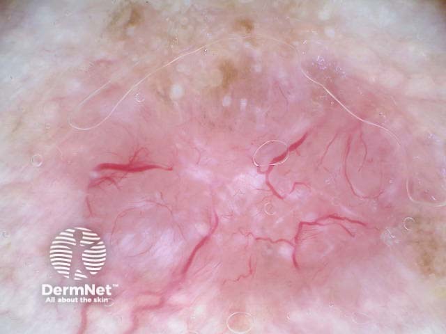
Melanoma in situ, polarised dermoscopy view
In which lesions are perpendicular white lines seen in through the dermatoscope?
Perpendicular white lines can be seen in the following lesions:
Pigmented and nonpigmented basal cell carcinoma
Melanoma
Spitz naevus
Dermatofibroma (the polarised white lines are often in radial array rather than perpendicular)
Scar tissue
Benignlichenoidkeratosis.
Perpendicular white lines in a variety of lesions
Superficial basal cell carcinoma dermoscopy
Nodular basal cell carcinoma dermoscopy
Polarised dermoscopy of a nodular basal cell carcinoma presenting as an exophytic polyp
Invasive melanoma dermoscopy, Breslow 0.4mm within a melanoma in situ, with associated naevus present
Amelanotic melanoma dermoscopy
Amelanotic melanoma dermoscopy
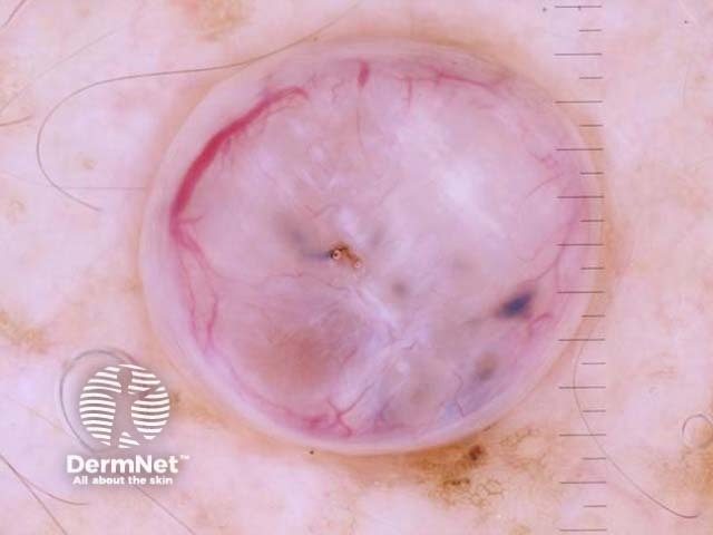
Spitz naevus dermoscopy
Dermatofibroma dermoscopy
Dermatofibroma dermoscopy
What is the histological explanation of perpendicular white lines?
Perpendicular white lines are thought to correlate histopathologically with altered collagen in the dermis (fibrosis). The birefringent properties of collagen bundles cause rapid randomisation of polarised light. This is the reason collagen appears bright white and is more conspicuous under polarised dermoscopy [3].
They also correlate with dermal invasion in cases of melanoma [4]. However, as our illustrations show, they may also be seen in melanoma in situ.
Bibliography
Kittler H, Marghoob AA, Argenziano G, et al. Standardization of terminology in dermoscopy/dermatoscopy: Results of the third consensus conference of the International Society of Dermoscopy. J Am Acad Dermatol. 2016 Jun;74(6):1093–106. doi: 10.1016/j.jaad.2015.12.038. Epub 2016 Feb 17. PMID: 26896294; PMCID: PMC5551974. PubMed Central
Balagula Y, Braun RP, Rabinovitz HS, et al. The significance of crystalline/chrysalis structures in the diagnosis of melanocytic and non-melanocytic lesions. J Am Acad Dermatol. 2012;67(2);194: e1–8.PubMed
Verzi et al.: The diagnostic value and histologic correlate of distinct patterns of shiny white streaks for the diagnosis of melanoma: A retrospective, case-control study. J Am Acad Dermatol. 2018;78:913–9. PMID: 29138058. Journal