Author(s): Dr Vera Miao, Northern Sydney Local Health District, Australia; Professor Saxon D Smith, Dermatologist, Sydney Adventist Hospital Clinical School, Australia (2023). Reviewing dermatologist: Dr Ian Coulson
Peristomal intestinal metaplasia (PIM) is a rare but serious complication affecting the skin around a gastrointestinal stoma site.
It is characterised by the histological appearance of scattered intestinal mucosa and goblet cells within the variably intact epidermis around the stoma site.
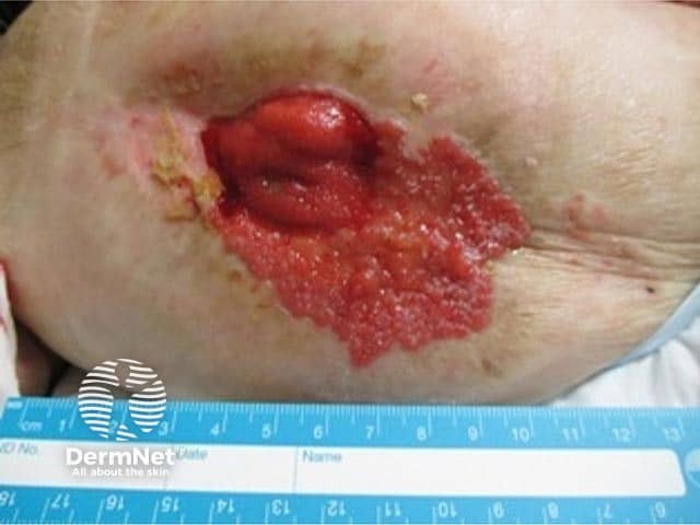
Well-definedgranulareruption emanating from ileostomy site (PIM-patient1)
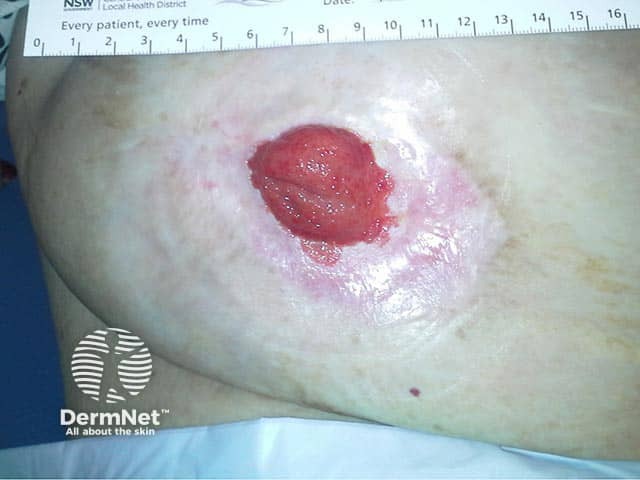
Re-epithelialised granular eruption which was emanating from ileostomy site (PIM-patient1)
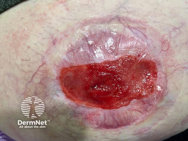
Well-defined granular eruption emanating from ileostomy site
Who gets peristomal intestinal metaplasia?
PIM is rarely reported and may occur in those with an ileostomy or a colostomy.
Chronicinflammation and irritation of the peristomal skin
Other peristomal skin conditions, most commonly peristomal dermatitis
Effluent leakage
Ill-fitting stomal appliances
Peristomal hernia.
What causes peristomal intestinal metaplasia?
The exact pathogenesis is not yet understood.
Suggested causative factors include:
Chronic inflammation of the peristomal skin
Exposure of the peristomal skin to intestinal bile salts in the stomal effluent, which increases intestinal epithelial cell migration
Seeding of intestinal mucosal cells into the peristomal skin during surgical procedures.
What are the clinical features of peristomal intestinal metaplasia?
PIM may present with the following clinical features:
Painful and irritated peristomal skin
Erosiveplaque/s around the stoma site
Peristomal ulceration
Peristomal maceration.
How do clinical features vary in differing types of skin?
Limited case reports in the literature make it difficult to compare presentations between different ethnicities. However, to date, there have been similar presentations across different Fitzpatrick skin types.
What are the complications of peristomal intestinal metaplasia?
Adenomas (polyps) in the peristomal skin
Malignant transformation to adenocarcinoma
How is peristomal intestinal metaplasia diagnosed?
The diagnosis of PIM is confirmed by skin biopsy, typically shave biopsy, of the epithelium at the peripheral border of the plaque/lesion. The characteristic histological appearance of PIM is that of intestinal mucosa infiltrating the epidermis around the stoma site.
What is the differential diagnosis for peristomal intestinal metaplasia?
What is the treatment for peristomal intestinal metaplasia?
General measures
Well-fitting stoma appliances — a specialist stoma nurse will advise on the most appropriate appliance and ensure the product adheres to the peristomal skin comfortably to minimise irritation and leakage.
Consider sequential electrocautery under local anaesthetic monthly for 4 months, and then as indicated for any sites of recurrence.
Revision of the stoma is sometimes found to be indicated — this is usually a collaborative decision involving the patient, surgeon, and dermatologist.
Serial monitoring for progression to adenocarcinoma.
In the event of adenocarcinoma developing in the metaplastic tissue, treatments may include:
- Surgical excision of the carcinoma
- Chemotherapy
- Radiotherapy
- Intralesional 5-fluorouracil (5FU)
How do you prevent peristomal intestinal metaplasia?
Proper stoma care and education by a trained professional, preferably a specialist stoma nurse.
Well-fitting stoma appliances to minimise effluent leakage and mechanical stripping of the peristomal skin.
Early recognition of the onset of the condition to allow for early intervention.
What is the outcome for peristomal intestinal metaplasia?
Electrocoagulation therapy has produced promising results in most case reports to date. Unfortunately, the rate of transformation to adenocarcinoma is not known.
Bibliography
Adachi K, Yamada N, Yoshida Y, Yamamoto O. Unusual features of peristomal erosion: a quiz. Intestinal epithelium on a peristomal erosion. Acta Derm Venereol 2010; 90: 223–244. doi: 10.2340/00015555-0755. Journal
Davey J, Day D, Howard V, Smith S. Peristomal intestinal metaplasia: A case report. Journal of the American Academy of Dermatology. 2016;74(5). doi: 10.1016/j.jaad.2016.02.393. Journal
Johnson CD, White H. Colonic metaplasia with colonic-type polyps on an ileostomy stoma in polyposis coli. Diseases of the Colon & Rectum. 1988;31(5):405-407. doi:10.1007/bf02564900. Abstract
Lyon CC, Smith AJ, Griffiths CE, Beck MH. The spectrum of skin disorders in abdominal stoma patients. British Journal of Dermatology. 2000;143(6):1248-1260. doi: 10.1046/j.1365-2133.2000.03896.x. Journal
Ono R, Oka M, Sakaguchi M, et al. Peristomal skin ulcer with intestinal metaplasia. British Journal of Dermatology. 2012;167(1):204-206. doi: 10.1111/j.1365-2133.2012.10819.x. Journal
Tao J, Joseph MX, Vaudreuil A, Dahiya M, Eilers D. Cutaneous Intestinal Metaplasia With Adenocarcinoma Treated With Intralesional Fluorouracil. JAMA Dermatol. 2023;159(1):111–112. doi:10.1001/jamadermatol.2022.4842 Journal
Tinker DG, Roberts S, Hurley MY, Missall TA. Cutaneous intestinal metaplasia: An unusual cause of peristomal complication with malignant potential. Journal of Cutaneous Pathology. 2020;47(5):479-480. doi: 10.1111/cup.13631. Abstract
Udechukwu NS, Selim MA, Nicholas MW. A case of periostomy intestinal metaplasia without adenomatous or dysplastic changes in an ulcerative colitis patient. Clinical Case Reports. 2020;8(3):535–7. doi: 10.1002/ccr3.2678. Journal