
ADVERTISEMENT
You do not have any notes added to this page yet
Introduction
Demographics
Causes
Clinical features
Variation in skin types
Diagnosis
Differential diagnoses
Treatment
Prevention
Outcome
Neurocutaneous melanosis (NCM) is a rare neurological disorder involving the abnormal accumulation of melanin within the central nervous system (CNS), accompanied by the presence of congenital melanocytic naevi (nevi).
NCM is thought to arise from the aberrant proliferation of neural crest-derived melanocytes. This leads to the development of benign or malignant melanocytic tumours within the CNS, along with at least one congenital melanocytic naevus (CMN).
Those with NCM may remain asymptomatic or show neurological signs and symptoms, most frequently seen in the first two years of life.
Neurocutaneous melanosis may also be referred to as neurocutaneous melanocytosis, neurocutaneous melanomatosis or neurocutaneous melanosis syndrome.
The prevalence of NCM is estimated at 1:50,000–1:200,000, with no clear gender predominance. Symptomatic NCM has been reported to manifest in approximately one-third to half of cases, typically in the first two years of life.
The estimated risk of NCM developing in individuals with large and giant congenital melanocytic naevus (CMN) is 10–33%.
Features of CMN that are associated with an increased risk of NCM include:
NCM is caused by an error within the neural crest cells of the ectoderm that results in the proliferation of mutated melanin-producing cells in the skin and leptomeninges.
In 2001, Takayama et al suggested this mutation within neural crest cells stemmed from atypical expression of hepatocyte growth factor/scatter factor.
Kinsler et al (2013) discovered another contributing factor to NCM, involving somatic mosaicism of a postzygotic mutation in codon 61 of NRAS, a developmental gene and oncogene playing a pivotal role in the regulation of crucial cell signalling pathways.
Building upon this research, Pawlikowski et al (2015) reasoned that activated Wnt signalling, in addition to the NRAS mutation, could contribute to the diverse phenotype observed in cases of NCM.
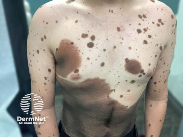
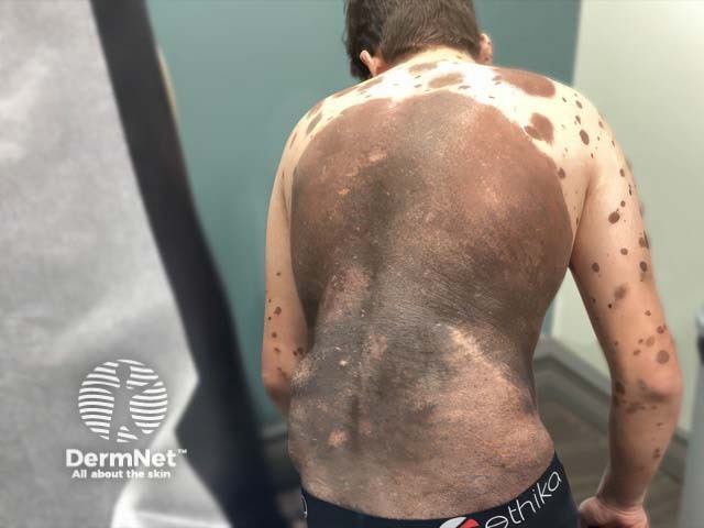
NCM tends to occur in patients with multiple medium-sized (1.5–20 cm) or large (>20–40 cm), or at least one giant (>40 cm) congenital melanocytic naevi (CMN). Note in children, these categorisations are based on projected size of the CMN in adulthood. Earlier size classifications differed with ‘giant’ CMN being previously considered >20 cm in diameter.
Multiple smaller naevi, sometimes referred to as satellite lesions, increase the risk of developing NCM.
NCM is also more likely in those with a ‘bathing trunk’ CMN distribution in the truncal, sacral, and perineal areas.
CMN may have associated hypertrichosis and verrucous-like texture and can develop papules and nodules.
Symptoms of neural melanosis, if they occur, may include manifestations of increased intracranial pressure that usually transpire within the first two years of life, such as:
Neurocutaneous melanosis can also cause stillbirth.
Dandy-Walker malformation (a rare congenital malformation of the cerebellum and fourth ventricle) has been observed in approximately 10% of individuals diagnosed with NCM. There also have been reports of NCM cases co-occurring with Chiari malformation, arachnoid cysts, choroid plexus papilloma, cerebellar astrocytoma, and spinal dysraphism, indicating a broader spectrum of associated conditions.
No specific data published on this.
Both skin and CNS lesions are at risk of malignant transformation, with leptomeningeal tumour transformation occurring in 40–60% of symptomatic cases. A giant CMN is associated with an estimated 3–8% lifetime risk of developing melanoma.
NCM can be further complicated by hydrocephalus, seizures, cranial nerve dysfunction, and the sequelae of spinal cord and root involvement.
In 1991, Kadonaga and Frieden revised the criteria for NCM to include:
Points 2 and 3 help to distinguish neurocutaneous melanosis from cases of metastatic melanoma eg, cutaneous melanoma that has metastasised to the CNS.
The preferred imaging modality is an MRI brain and spine with the recommendation of prompt evaluation, ideally within the first 4–6 months of life, for patients with one or more of the following features:
Solitary small, medium, and large CMN are generally considered low risk for NCM, therefore MRI brain and spine may not be indicated in these patients. There is debate around this and some suggest imaging all patients with large CMN (>20 cm) or >1 CMN.
MRI appears to be most sensitive to melanosis in the first few months of life and is the best test to evaluate the involvement of the leptomeninges. Non-contrast MRI may be attempted on young infants using a ‘feed and swaddle’ technique that avoids the need for general anaesthetic.
Melanocyte deposition appears hyperintense on T1-weighted MRI due to its paramagnetic properties, with focal shortening of T1 relaxation time compared with T2. Neural melanosis is most commonly seen in the amygdala, and sometimes the inferior cerebellum, ventral medulla, pons, cerebral peduncles, or the upper cervical spinal cord. Deep parenchymal structures are usually spared.
MRI findings for malignant CNS lesions may include a mass with nodular or thick plaque-like contrast enhancement, oedema, rapid growth, necrosis, or haemorrhage. However, MRI alone often cannot distinguish benign from malignant neural melanosis.
Cerebrospinal fluid (CSF) analysis and the use of ultrasound have been reported. Transfontanellar ultrasonography may reveal increased echogenicity of the amygdala if lesions are present and can aid in the detection and monitoring of hydrocephalus.
Depending on MRI brain and spine findings, differential diagnoses could potentially include:
Specialist dermatologist follow-up is important. All children with a confirmed diagnosis of neurocutaneous melanosis should also be referred to a paediatric neurologist. Neurosurgical input may also be needed.
It is recommended to conduct lifelong monitoring, including visual inspection +/- dermoscopy and palpation of each CMN every 6–12 months, and palpation for lymphadenopathy. Baseline and serial photographs and detailed mole-mapping can help track any alterations. Any palpable nodules or changes such as rapid growth or new induration or ulceration warrant consideration of histological examination. More frequent visits may be recommended during infancy and puberty, which are times of increased naevus change and growth.
It may be reasonable to contemplate surgical excision / removal of CMN in children ≥6 months of age. However, CMN removal does not fully protect against the onset of melanoma as half of these are derived from melanocytes elsewhere in the body (eg, CNS). Careful consideration of risks and benefits of CMN removal is needed, including factors such as the size and location of the naevus, patient and/or family preference, and the overall prognosis.
There is no definitive treatment for NCM, and chemotherapy and radiation therapies are not effective in modifying the disease course.
Therapies to help manage neurological symptoms associated with the disorder include ventriculoperitoneal shunts to alleviate intracranial pressure in cases of hydrocephalus, and anticonvulsant medications to reduce seizures.
New targeted therapies are being explored, including inhibitors specific for Kit-ligand, hepatocyte growth factor, or the MET signalling pathway.
Drugs that block the MAPK (mitogen-activated protein kinase) pathway through MEK inhibition in cases of NCM have shown potential promising effects.
While there are no specific preventive measures for NCM, recommended screening protocols for patients with congenital melanocytic naevi can play a pivotal role in early detection and improved monitoring of the condition. In patients with neurological symptoms or asymptomatic patients with at least one risk factor (eg, multiple satellite lesions; and/or a large-sized CMN, especially on the posterior axis), an MRI should be considered prior to 4–6 months of age for the highest sensitivity.
Prognosis for patients with neurological signs and symptoms is much worse than for those without. Those that remain asymptomatic have a normal life expectancy.
More than 50% of symptomatic patients will pass away within three years of symptom onset.
The mortality rate of melanoma arising in the CNS is almost 100%.
Brock Elbank’s congenital melanocytic naevus (CMN) photography series: HOW DO YOU C ME NOW? — Hasselblad
An AI summary will appear based on your search term using data from all of the topic pages across the entire DermNet site.
Show more