Necrotising fasciitis is a life-threatening, rapidly progressive form of necrotising cellulitis. Group A beta-haemolytic streptococci are the most common causative organisms. Various other bacteria have been implicated.
Histology of necrotising fasciitis
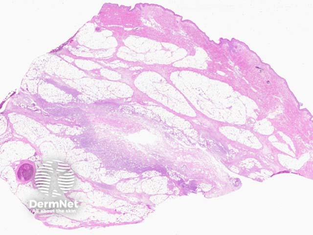
A deep biopsy is generally required (figure 1) for the diagnosis and shows an extensive acuteinflammatory reaction involving the subcutaneous fat (figure 2). There may also be superficial involvement and ulceration and/or deep penetration into skeletal muscle and deeper tissues.
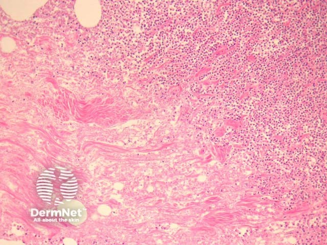
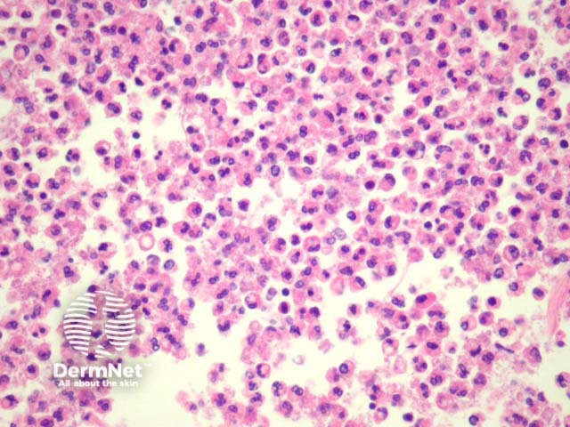
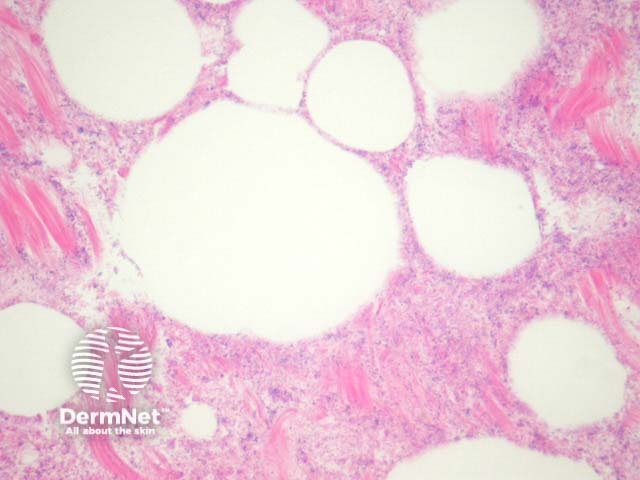
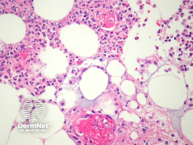
Higher power examination demonstrates impressive suppuration (figure 3) and extensive tissue necrosis (figure 4). An overwhelming bacterial colonisation is appreciated even on haematoxylin-eosin sections (figure 4). There is often associated intravascularthrombosis (figure 5) and invasion of vessel walls by micro-organisms.
Figure 1
Figure 2
Figure 3
Figure 4
Figure 5
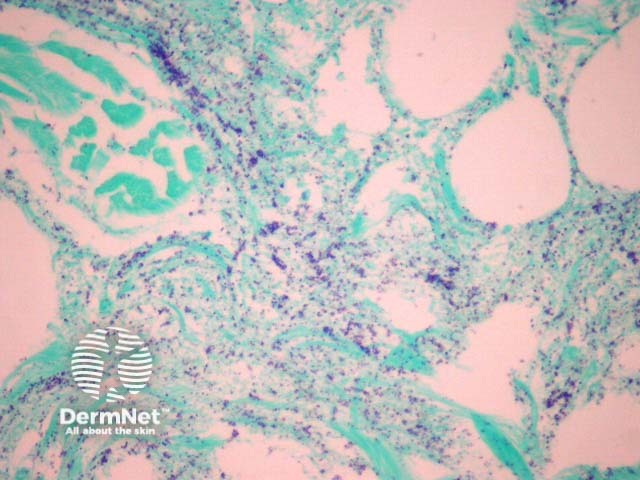
Figure 6
Special studies for necrotising fasciitis
Gram stain confirms the overwhelming colonisation by the micro-organism (figure 6). The case illustrated shows numerous gram-positive cocci but the morphology and staining characteristics will be varied depending on the organism involved. Tissue culture confirms the causative organism.
Intraoperative consultation for frozen section is sometimes employed for this disease. In addition to routine frozen section assessment, touch preparations can be useful for identifying the presence of an organism. Correct diagnosis is critical for appropriate surgical management of this aggressive disease.
Differential diagnosis of necrotising fasciitis pathology