Edit profile
You do not have any notes added to this page yet
Topics A-Z
Mohs surgery images
Treatments
Author: Tom Middelburg, MD PhD, dermatologist and Mohs surgeon, Christchurch, New Zealand, May 2017.
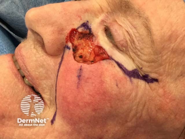
Example of a Mohs procedure
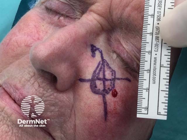
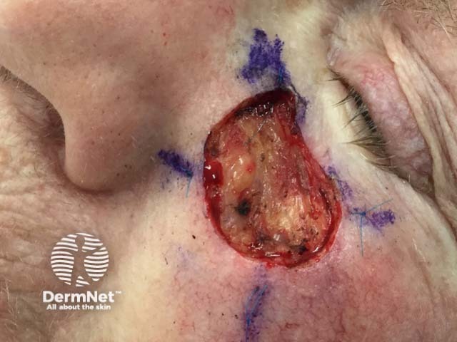
This infiltrative basal cell carcinoma required 5 Mohs stages.
Clinically visible basal cell carcinoma plus margin is outlined, a grid is created as part of the ma
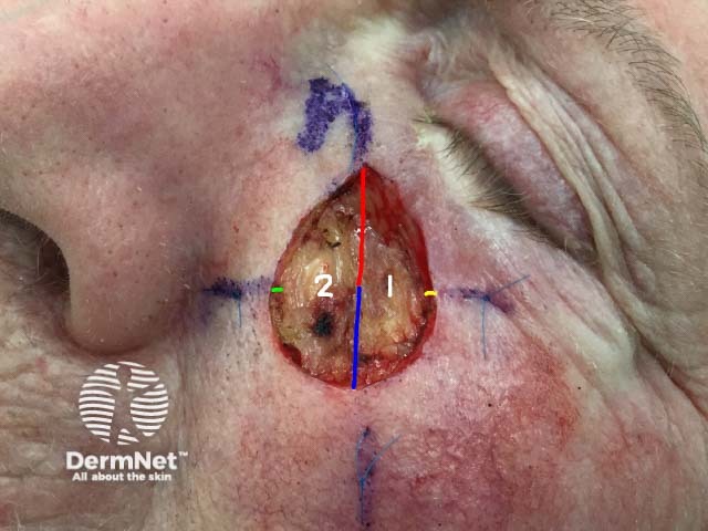
Tissue sample is excised and cut into two specimens which are colour code
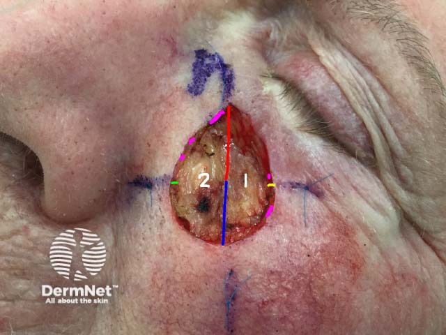
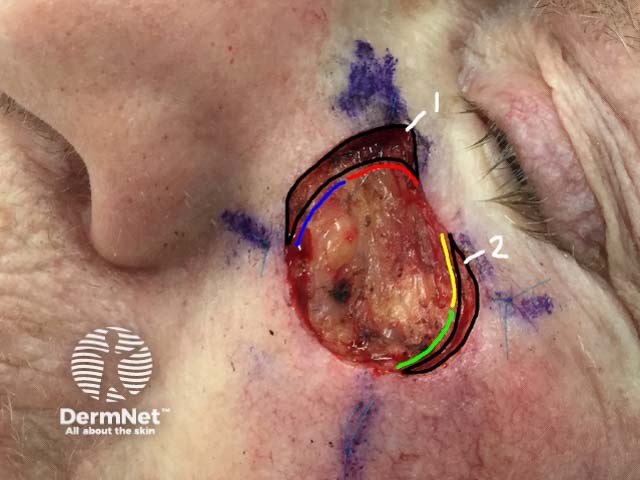
After microscopic examination of histological slides any areas of residual tumor are recognised and
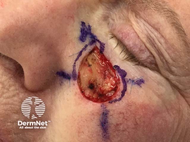
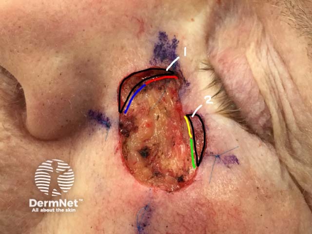
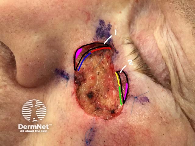
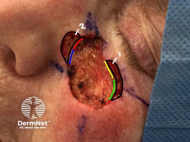
In the second Mohs stage the area to be excised is marked on the patient
The marked areas are excised
The two tissue samples are colour coded
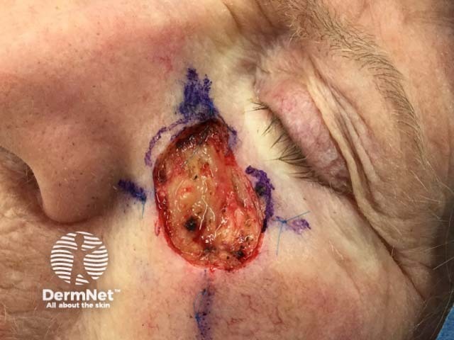
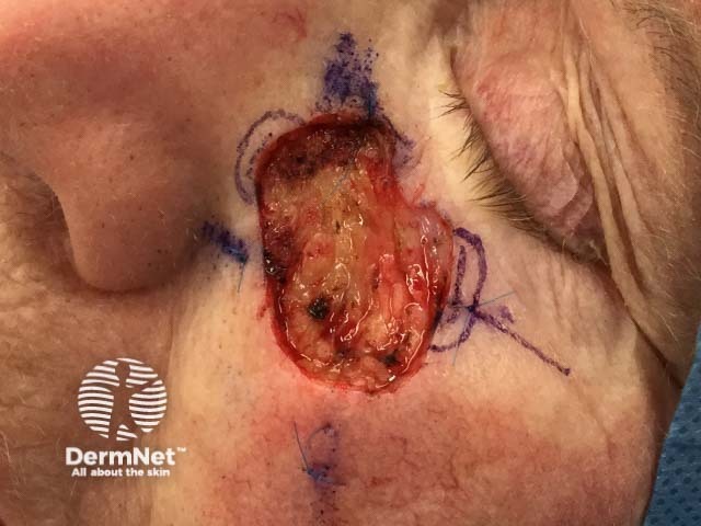
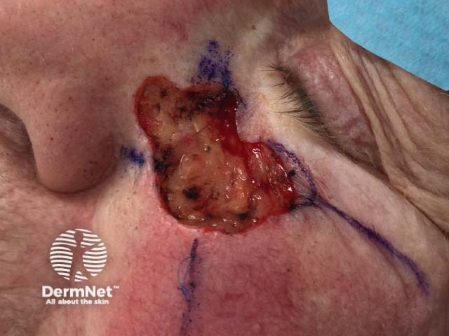
In the third Mohs stage the area to be excised is marked on the patient
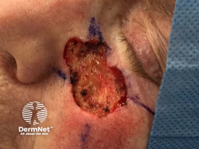
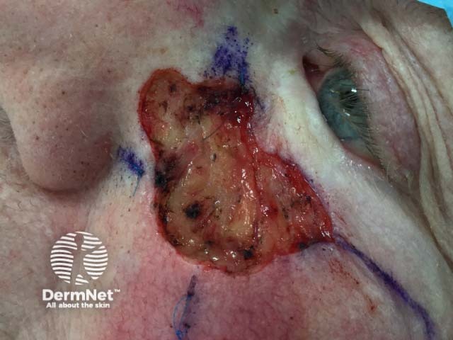
In the fourth Mohs stage the area to be excised is marked on the patient
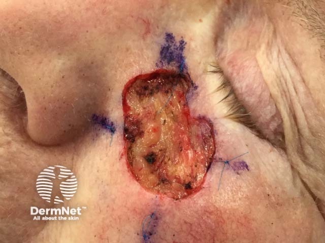
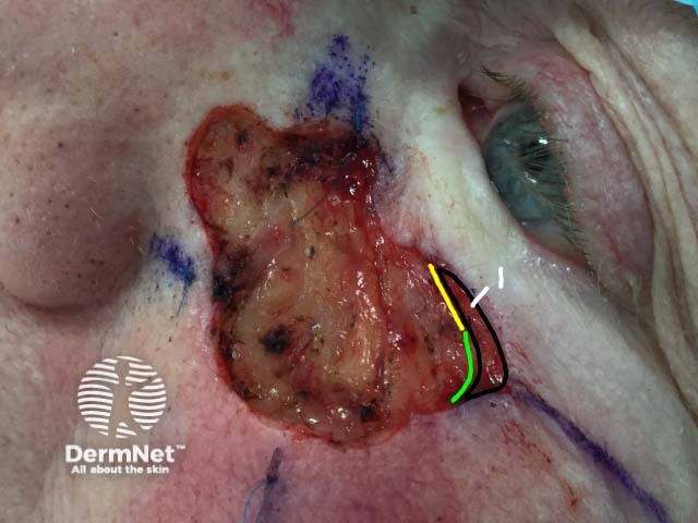
In the fifth Mohs stage the area to be excised is marked on the patient
The marked area is excised
The area is free of tumour cells
The wound is reconstructed, in this case by a cheek advancement flap
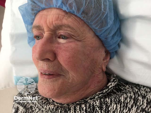
Two months after the Mohs procedure
Showing 0 of 0 conditions
Advanced filters
Reset filters
An AI summary will appear based on your search term using data from all of the topic pages across the entire DermNet site.
Show more