
ADVERTISEMENT
You do not have any notes added to this page yet
Introduction
Clinical features
Causes
Investigations
Treatment
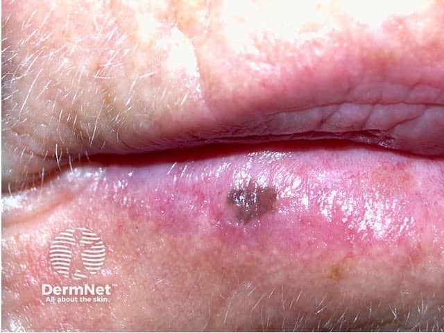
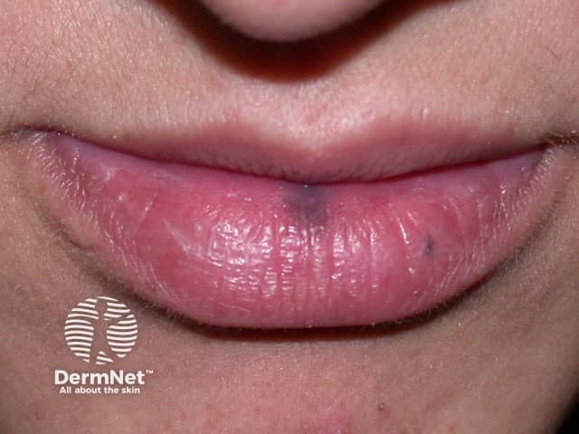
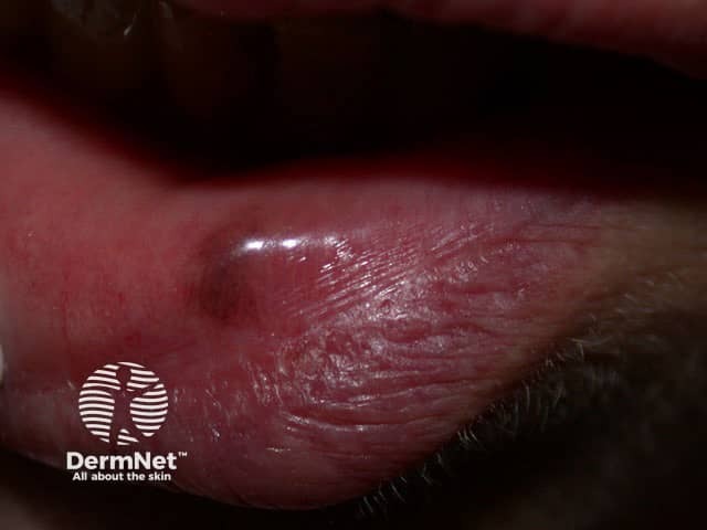
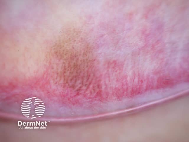
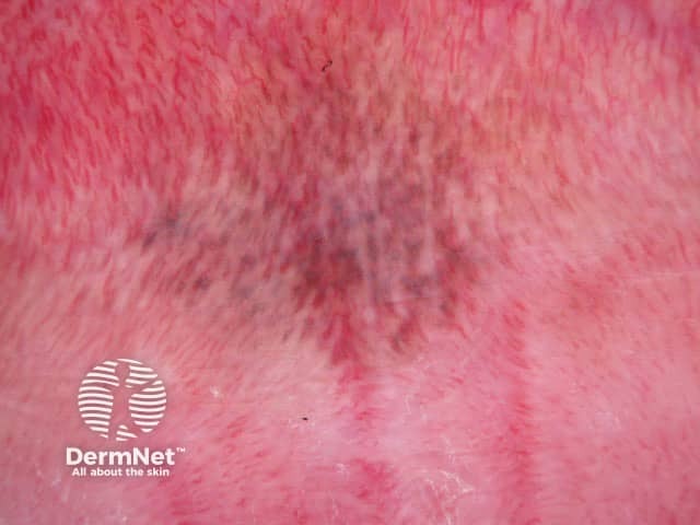
A melanotic macule is a well-defined, oval, brown to black, flat patch usually found on the central third of the lower lip, however it can more rarely occur on the penis or vulva.
A labial melanotic macule is the name for a freckle arising on the lip. It is also sometimes called a labial lentigo and when multiple lesions are present, mucosal melanosis.
Usually solitary, a labial melanotic macule is most commonly seen in adult women but it also occurs in males and in young people. Occasionally the lesion can be on the upper lip.
Size ranges from 1–8 mm. Once developed, the lesions usually remain unchanged in size and colour. They can occasionally have an irregular edge and there may be a history of colour change which can cause confusion with other pigmented lesions, including melanoma. Luckily, melanoma is very rare on the lip (but it can occur).
Similar freckles may also occur in areas that are not exposed to the sun:
Labial melanotic macules do not cause any symptoms but their appearance can be a concern to the patient.
A labial melanotic macule is thought to be provoked by sun exposure, and it is more common in fair-skinned people. However, it may also occur in dark-skinned individuals and, as described above, similar lesions can arise in sites that are never sun-exposed. Luckily, melanotic macules are harmless.
A labial melanotic macule may be confused with another pigmented skin lesion.
These conditions can be differentiated from labial melanotic macule by a combination of clinical and histological features.
Multiple lesions may be a sign of a widespread skin condition, such as:
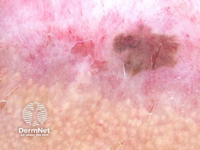
A melanotic macule has a characteristic pattern when examined with the magnifying glass or by dermoscopy. Lesions with a typical history and appearance need not be biopsied.
If a skin biopsy is done because the lesion is changing or looks irregular, a labial melanotic macule shows the following features on dermatopathology:
Nuclear atypia is absent and the melanocyte count is normal.
Typical lesions can just be observed. Suspicious lesions, including lesions showing progressive change, should be biopsied.
If treatment is requested the macules can be frozen (cryotherapy) or removed using a laser or intense pulsed light. Excision can also be performed but will leave a scar.
An AI summary will appear based on your search term using data from all of the topic pages across the entire DermNet site.
Show more