These guidelines were provided to DermNet by ProCare Health Limited, July 2014
Disclaimer
These guidelines have been written for the use of ProCare member practices. No set of guidelines can cover all variations required for specific patient circumstances. It is the responsibility of the health care practitioners using these guidelines to adapt them for safe use within their institutions and for the individual needs of patients.
Definition
Cryotherapy is a minimally invasive procedure that uses an extremely cold liquid or instrument to freeze and destroy abnormal tissue that requires elimination. It is also referred to as cryosurgery or cryoablation.
Scope
Medical practitioners and registered nurses who have been are trained to perform the procedure.
Treatment of malignant skin lesions by cryotherapy is not covered by this document.
Indications for cryotherapy
Viral warts in older children and adults
Seborrhoeickeratoses
Actinic keratoses
Molluscum contagiosum in adults
Skin tags*
*Diathermy may be more effective for acrochordons / fibroepithelial polyps
The following skin cancers may be suitable for cryotherapy if performed by a medical practitioner with appropriate training and where the lesion has been identified by biopsy:
Small, thin, typical, superficial basal cell carcinoma on trunk and limbs
Small, typical intraepithelial squamous cell carcinoma on trunk and limbs
Contraindications to cryotherapy
Undiagnosed skin lesions
Lesion for which tissue pathology is required
Lesion within a circulation compromised area
Melanoma
Previous sensitivity or adverse reaction to cryosurgery
Patient unable to accept side effects
Patients with poor circulation
Unconscious patients
Young children
Dark skinned patients
Precautions when using cryotherapy
Areas not recommended for liquid nitrogen application: corners of eyes, fold of skin between nose and lip, skin surrounding nostrils and skin overlying nerves, e.g. sides of digits, below the knee in certain groups (eg diabetics, elderly)
Re-appearance of a lesion previously treated with cryotherapy should be referred for review by medical practitioner
Recurrent skin cancers after cryotherapy may be more difficult to treat
Exercise care in patients with history of slow healing or skin infection
Prolonged freezing may result in scarring – better to freeze lightly and for the patient to return for re-freeze if response is inadequate
Cryotherapy leaves permanent white marks which may be very unsightly, especially in dark skinned patients
Cryotherapy may sometimes cause nerve damage and on-going pain in some danger areas where the nerves lie superficially (eg sides of the fingers)
Checklist for cryotherapy
Pre-procedure
General practitioner should review any lesion where there is uncertainty about the diagnosis or suitability for cryotherapy
Obtain informed consent
Prepare equipment/environment
Apply protective eyewear and gloves
Decant liquid nitrogen into the cryospray or if using a cotton tipped applicator into a non-permeable container
Cotton tipped applicators have been commonly used but they should only be used for benign lesions, owing to inferior tissue freezing compared to spray techniques
Large cotton swabs used for cryotherapy
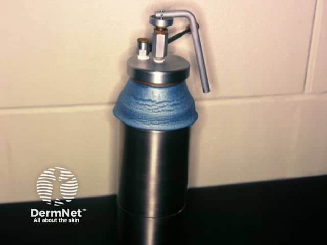
Cryotherapy liquid nitrogen dispenser
Liquid Nitrogen Application
Hand hygiene
Select spray tip A to D of the appropriate size for the diameter and thickness of the lesion
Apply the liquid nitrogen to the lesion for a few seconds, depending on the required diameter and depth of the freeze
A margin of 1–2 mm is recommended for benign lesions
A disposable plastic ear speculum (trimmed if necessary) can be used to confine the area of treatment
Freeze times vary from around five seconds (after the freeze ball appears) for actinic keratoses to 10 or 20 seconds for thicker lesions such as plantar warts or seborrhoeic keratosis
Two freeze/thaw cycles (with a shorter freeze time) are more effective for thicker lesions such as seborrhoeic keratosis and warts
For warts (especially plantar warts) removal of keratin by the use of a scalpel blade or prior keratolytic treatment (eg salicylic acid) may improve the response to subsequent cryotherapy
Cotton tipped applicators should not be re-dipped into the flask; new swabs should be used if more liquid nitrogen is required
Post–procedure
Periodic cleaning and sterilisation of cryospray nozzles should be performed according to manufacturer's recommendations
If the nozzle comes in contact with patient skin then sterilisation by autoclave is required (refer to the practice Infection Control policy)
Wipe down flask
Document the lesion and treatment in the patient management system. This includes (but not limited to)
Informed consent obtained
Specific instructions from the general practitioner
Lesion site
Duration of liquid nitrogen application
Follow-up advice
Inform patient that the treated area:
May blister within a few hours (clear, red or purple)
Bleeding may occur (though this is not common)
The blister shrinks to be replaced by a scab within a few days
Swelling should settle in a few days
Healing depends on the site the scab peels off within a week after cryotherapy to facial actinic keratoses, after about three weeks to a similar lesion on the hand, and may cause ulceration on the lower leg and take three months or longer to heal
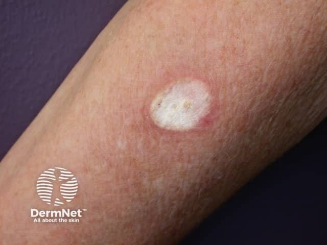
A white mark (hypopigmentation) or scar may result
If there are any signs of infection the patient should contact the practice**
**For example, increasing redness of surrounding skin, discharge, increasing pain
Frozen skin
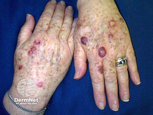
Blistering one day post liquid nitrogen application
Complications and side-effects
Acute
Oedema
Pain
Headache after treatment of facial lesions
Delayed
Bleeding at the frozen site
Infection at the site
Skin discomfort
Hyperpigmentation (slowly resolves)
Permanent (uncommon)
Alteration of sensation
Hypopigmentation
Hypertrophic scarring
Hair loss
Safety considerations
Always ensure the working area has adequate ventilation when handling liquid nitrogen
Personal protection clothing, including leather gloves, safety glasses and covered footwear, are used when decanting the liquid nitrogen from the storage Dewar flask (storage container) to the cryospray or non-permeable container
Caution is to be taken when transporting liquid nitrogen
Liquid nitrogen Dewar flask and cryospray are to be stored in an upright position, in a cool, well-ventilated area away from heavily trafficked areas. These are to be secured to prevent accidental knocking
To eliminate the potential build of condensation liquid nitrogen dewar flask and cryospray should be stored closed as per manufacturers instruction
Dewar flasks used for liquid nitrogen storage must have a loose lid or ventilation in the lid to prevent build-up of pressure and consequent risk of explosion
Relevant practice policies and procedures
Health and Safety Policy
Informed Consent Policy
Infection Control Policy
Practice Hazard Register
Material Data Safety Sheets: liquid nitrogen
References
Aaron J. Morgan, Elston, M.D. – Medscape, Drugs, Conditions and Procedures, 2010
Cryotherapy – British Association of Dermatologists
Zimmerman EE, Crawford P. Cutaneous cryosurgery. Am Fam Physician. 2012 Dec 15;86(12):1118-24. Journal
Guidelines of care for cryosurgery. American Academy of Dermatology Committee on Guidelines of Care. J Am Acad Dermatol. 1994 Oct;31(4):648–53. PubMed
Miller-Keane Encyclopaedia and Dictionary of Medicine, Nursing, Allied Health, Seventh Edition, 2003 by Saunders
Royal New Zealand College of General Practitioners — The Standard for New Zealand General Practice, 2011–2014, Indicator 18, Medical Equipment