Infantile digital fibroma, also called inclusion body fibromatosis or Reye tumour, is a benignproliferation of myofibroblasts.
Histology of infantile digital fibromatosis
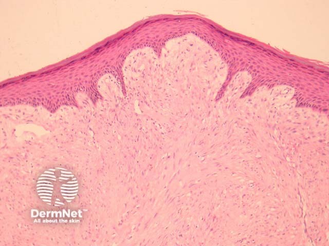
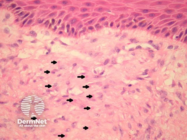
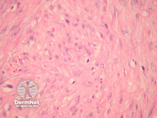
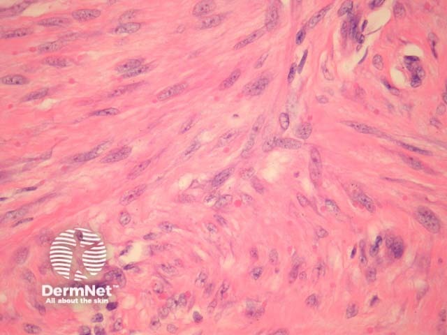
In infantile digital fibromatosis, sections show an intradermal unencapsulated tumour composed of spindle-shaped cells arranged in whorls or interdigitating sheets (figure 1). These myofibroblasts contain 3–10 μm inclusion bodies, which are round or ovoid and granular (figures 2-4, arrows are used to highlight some of the inclusion bodies in figure 2).
Earlier lesions of infantile digital fibromatosis are more inflammatory; more developed lesions display more fibroplasia and inclusion bodies.
Figure 1
Figure 2
Figure 3
Figure 4
Special studies for infantile digital fibromatosis
The inclusion bodies stain pink with H&E (figures 2-4). The bodies are positive with immunohistochemical stains for actin and vimentin.
Differential diagnosis of infantile digital fibromatosis pathology
Fibromatosis – Identification of the characteristic inclusion bodies distinguish infantile digital fibromatosis from dermal fibromatosis and hypertrophic dermal scars
References
Grenier N, Liang C, Capaldi L, Ney A, Lapidus C, Schappell D, Katarincic J, Robinson-Bostom L. A range of histologic findings in infantile digital fibromatosis. Pediatr Dermatol. 2008 Jan-Feb;25(1):72–5. PubMed