Impetigo is a common superficial bacterialinfection of the skin. Streptococcus and/or Staphylococcus species are the usual causative organisms.
Histology of impetigo
There are two clinical forms: bullous and common. The common type is rarely biopsied.
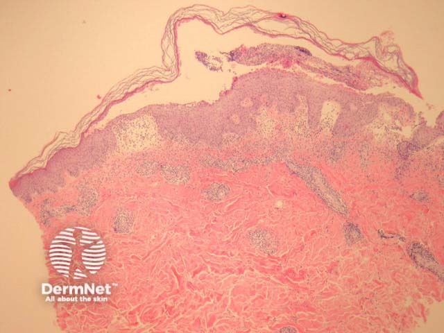
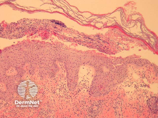
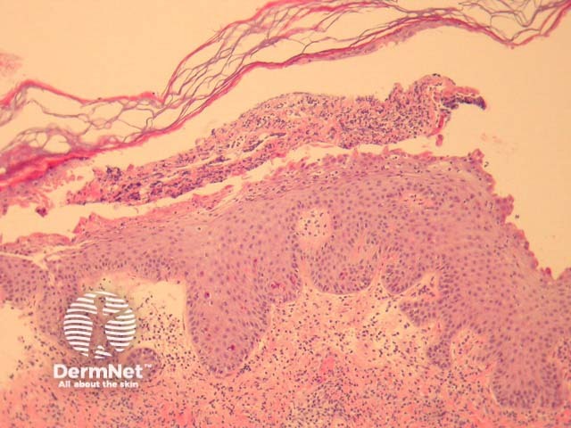
The bullous form of impetigo shows a subcornealbulla with a dermalinflammatory response (figure 1). There are acantholytic cells in the superficial epidermis accompanying degenerated keratinocytes and small numbers of neutrophils (figure 2, 3). Gram positive cocci may be seen within the superficial epidermis.
The common type of impetigo shows subcorneal collections of neutrophils, serum and parakeratotic foci. There is usually a dermal inflammatory response. The organisms may be difficult to isolate with gram stains.
Figure 1
Figure 2
Figure 3
Special studies for impetigo
Gram stain may be helpful to demonstrate the organisms. Correlation with culture results can sometimes be helpful.
Differential diagnosis of impetigo pathology
Staphylococcal scalded skin syndrome — also exhibits a subcorneal bulla with acantholytic cells. The inflammatory response in the dermis and superficial neutrophils are typically absent.
Pemphigus foliaceus — also exhibits a subcorneal bulla with acantholytic cells. The inflammatory response in the dermis and superficial neutrophils are typically absent. Immunofluorescence can be helpful in confirming this diagnosis.
References
Weedon’s Skin Pathology (Third edition, 2010). David Weedon
Pathology of the Skin (Fourth edition, 2012). McKee PH, J. Calonje JE, Granter SR