In dermoscopy, a grey circle consists of a continuous curved grey line, equidistant from a fixed centre point.
What do grey circles look like through the dermatoscope?
Grey circles are seen through the dermatoscope as a thin circular line of grey pigment.
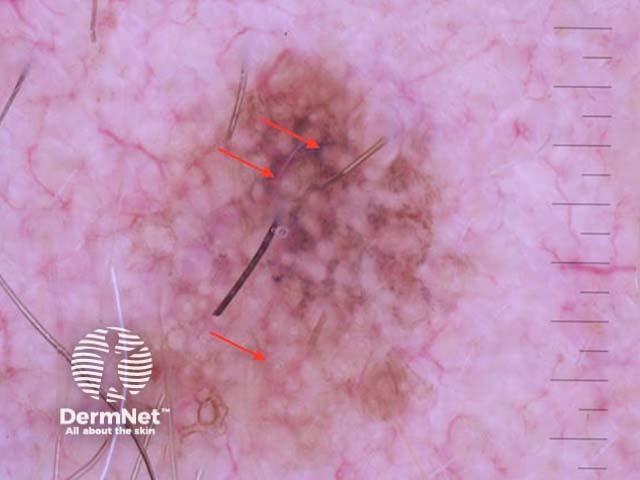
Grey concentric circles seen on dermoscopy of lentigo maligna
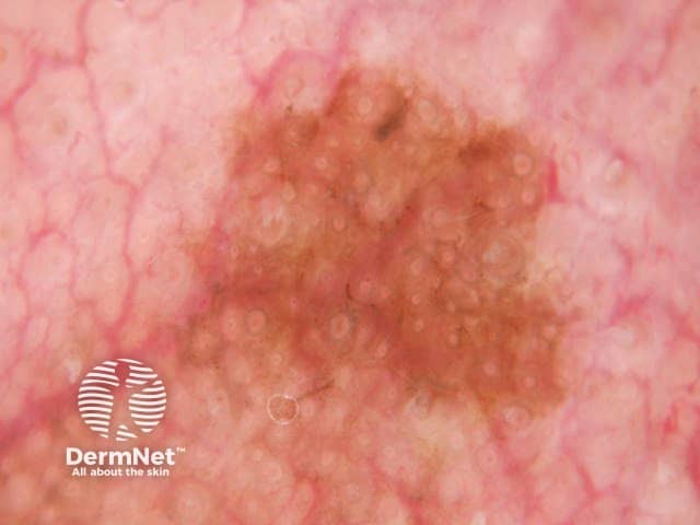
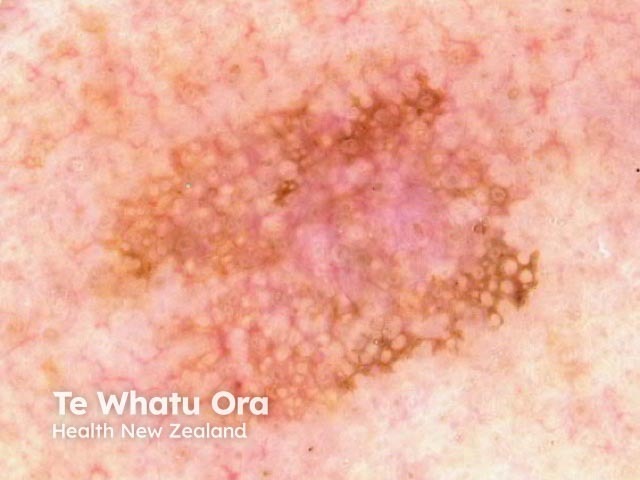
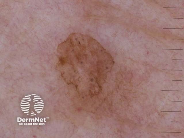
Grey circles in lentigo maligna
Grey circles in lentigo maligna
In which lesions are grey circles seen through the dermatoscope?
Dermoscopic grey circles are seen in the following lesions:
Solar lentigo
Pigmentedactinic keratosis
Pigmented intraepidermalcarcinoma
Lichen planus-like keratosis (LPLK)
Lentigo maligna.
Grey dots around the hairfollicle openings forming grey circles tend to be coarse in LPLK, whereas in lentigo maligna they are much finer.
A pattern of thin grey circles is the most specific clue to early facial melanoma, but only when pigmentation is confluent, and not grey dots arranged as circles [1]. Grey circles had a sensitivity of 54.2% and a specificity of 83.3% for melanoma on the face [2].
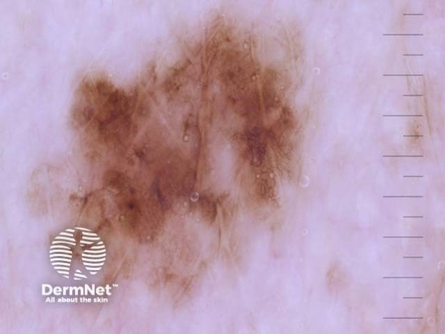
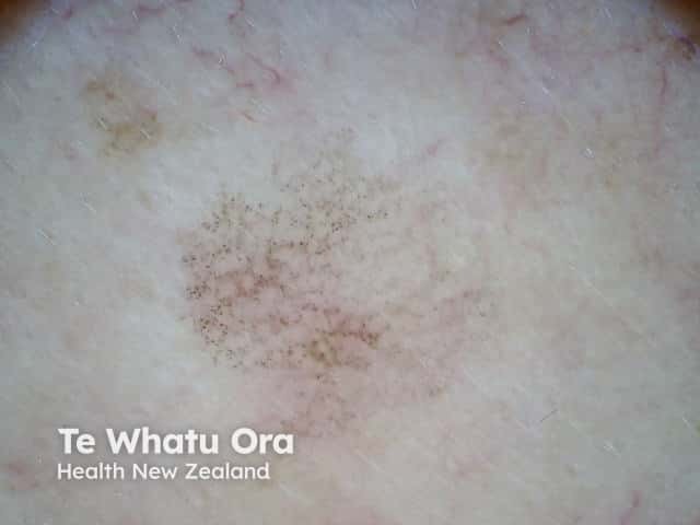
Grey circles seen in dermoscopy of a facial solar lentigo
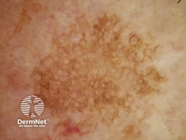
Grey circles in pigmented actinic keratosis
Grey circles in pigmented intraepidermal carcinoma
Annulargranular pattern seen in dermoscopy of lichen planus-like keratosis
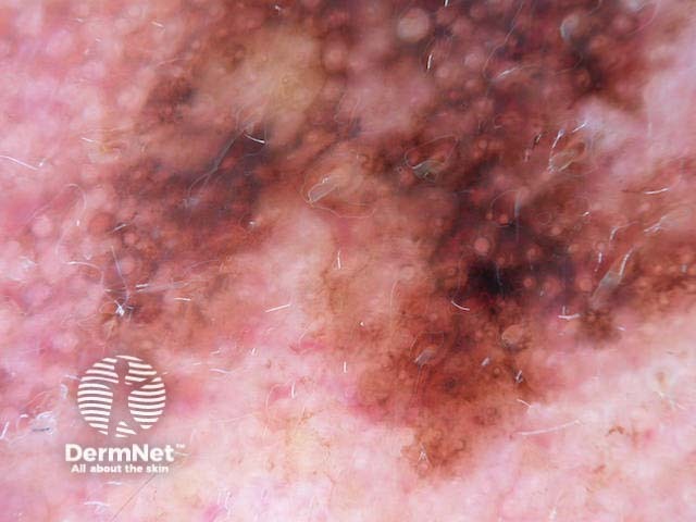
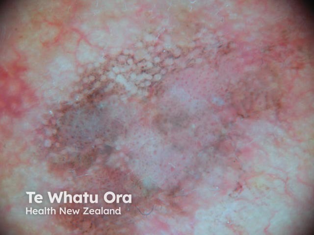
Grey circles in lentigo maligna
Grey circles in a solar lentigo
Circles within circles
A circle within a circle consists of an asymmetrical pigmented follicular opening with a darker dot located within the ostial opening [3]. Circles within circles are a specific clue for facial melanoma in situ but are rarely present; they have poor sensitivity and are found in only 4.2% of flat facial melanomas [2]. They can also be found in pigmented actinic keratosis, but rarely [3]. Circles within circles are also known as concentric circles and the 'isobar' sign.
Grey concentric circles in lentigo maligna
What is the histological explanation of grey circles?
Circles on facial skin reflect prominent hair follicles with associated pigmentation in an otherwise featureless epidermis. Pigmentation may be within the epithelium giving grey circles in melanophages around hair follicles resulting in circles comprising grey dots. Th may also involve the intervening epidermis giving grey circles in a structureless brown background. It should be noted that nonpigmented follicular openings do not constitute a pattern of circles [4].
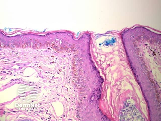
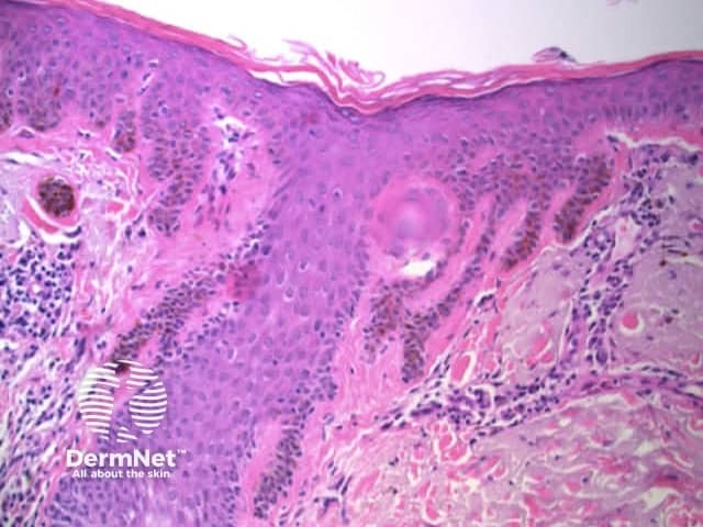
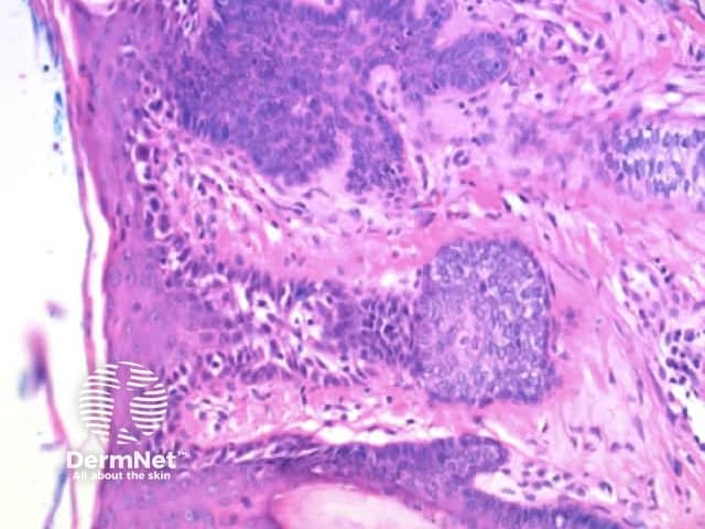
Grey circles correspond to pigmented melanocytes within follicular infundibula
Prominent perifollicular melanophages are seen as grey dotted circles
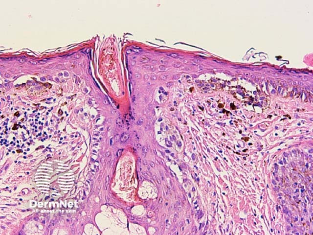
Involvement of follicular infundibula displayed as circles on dermatoscopy
Involvement of follicular infundibula displays as circles on dermoscopy
References
Kittler H, Rosendahl C, Cameron A, Tschandl P. Dermatoscopy, Pattern analysis of pigmented and nonpigmented lesions.. 2016. Facultas Verlags and Buchhandels AG, Universitatsverlag, Austria.
Tschandl P, Rosendahl C, Kittler H. Dermatoscopy of flat pigmented facial lesions. J Eur Acad Dermatol Venereol. 2015 Jan;29(1):120-7. doi: 10.1111/jdv.12483. Epub 2014 Mar 24. PubMed PMID: 24661420.
Florentia Dimitriou, Theresa Deinlein, Iris Zalaudek, 'Actinic keratosis', dermoscopedia, 6 June 2019, 12:56 UTC, https://dermoscopedia.org/actinic keratosis (accessed 5 August 2019).
Kittler H, Rosendahl C, Cameron A, Tschandl P. Dermatoscopy. An algorithmic method based on pattern analysis. 2nd ed. 2016. Facultas Verlags and Buchhandels AG, Universitatsverlag, Austria.