Extramammary Paget disease of the skin is a malignancy-associated skin disease characterised by an eczematousrash of the anogenital region and vulva, where there is an abundance of apocrine glands.Primarycutaneous forms exist associated with underlying adnexalcarcinoma, in addition to forms associated with underlying anal, rectal or bladder adenocarcinoma. Extramammary Paget disease may precede the associated malignancy by up to 15 years.
Histology of extramammary Paget disease
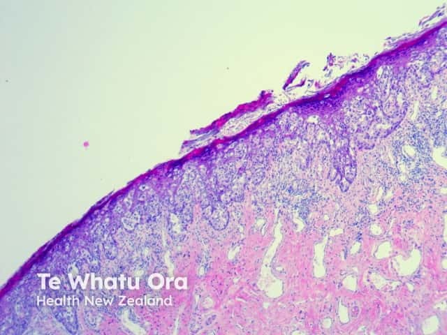
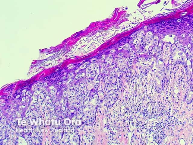
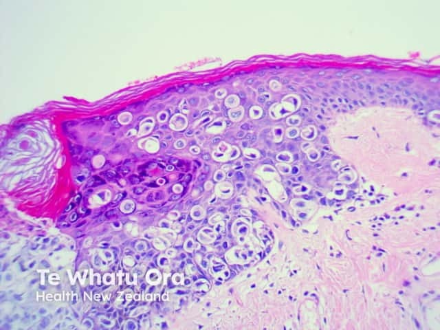
Paget tumour cells are located in the epidermis (Figure 1–3). Rarely, tumour cells can extend into the dermis. In earlier lesions, the Paget cells are located near to the basement membrane, and they extend to fill the entire epidermis as the condition progresses. Tumour cells are large with clear or eosinophiliccytoplasm, large pleomorphicnuclei and occasional mitoses (Figure 3). Hyperkeratosis and parakeratosis may overlie aggregates of tumour cells (Figure 2). Hyperplasia of the epidermis may be squamous, papillomatous or fibroepithelioma of Pinkus-like. Ulceration and pigmentation may be present. Dermal changes include a chronicinflammatoryinfiltrate (Figure 1, 2). An underlying adnexal carcinoma may be present.
Figure 1
Figure 2
Figure 3
Special studies of extramammary Paget disease
Tumour cells contain abundant mucin which can be identified by staining with Alcian blue and periodic acid-Schiff (PAS). Immunohistochemically, Paget cells stain for epithelial membrane antigen (EMA), low molecular weight cytokeratins (CK7+) and CD23 with variable positivity for carcinoembryonic antigen (CEA). High molecular weight cytokeratins stain the surrounding epidermis and highlight the negative Paget cells.
Differential diagnosis of extramammary Paget disease
Mammary Paget disease of the skin: this condition is a cutaneous lesion arising from an underlying ductal in-situ carcinoma of the breast. Extramammary Paget disease lacks the Her-2/neu expression seen in mammary Paget disease. Breast tumours and tumours originating from apocrineglands stain with GCDFP-15.
Melanomain situ — in contrast to Paget, melanoma is negative for cytokeratins, EMA and CEA. S100, HMB-45 and MART-1 are usually negative in Paget’s disease and positive in melanoma.
Squamous cell carcinoma (SCC) in situ (intraepidermal carcinoma) — P63 is positive in SCC in situ, differentiating from Paget. Cytokeratin seven may be positive in both Paget and SCC in situ.
Sebaceous carcinoma in situ — adipophilin positive, CEA negative, GCDFP-15 negative.