Epithelioid cell histiocytoma is a well recognised variant of cutaneous fibrous histiocytoma that may be confused with other benign and malignant mesenchymal lesions. It is otherwise known as epithelioid fibrous histiocytoma and epithelioid dermatofibroma.
Histology of epithelioid histiocytoma
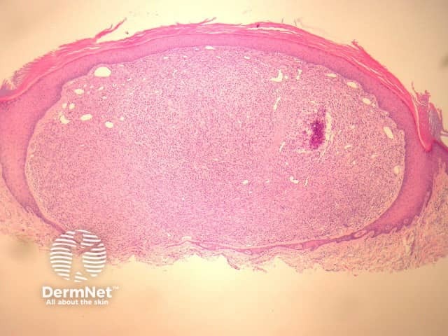
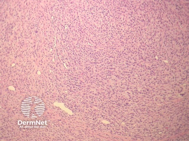
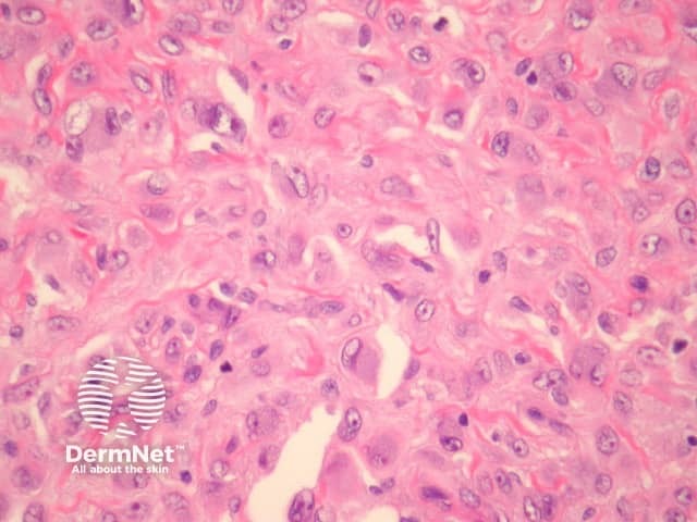
In epithelioid histiocytoma, sections show a centrally located, circumscribedtumour underlying an epidermal collarette (figure 1). The tumour is composed of epithelioid cells arranged in sheets and sometimes a storiform pattern. Individual cells show abundant eosinophiliccytoplasm with round vesicularnuclei and prominent nucleoli (figures 2-4). The cells may be markedly enlarged and display some nuclearatypia (figure 4).
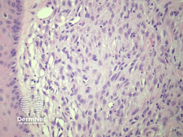
There is frequently an associated inflammatory cell infiltrate which can be helpful for the diagnosis (figure 3). Some mitoses may be seen. Multinucleatedgiant cells and haemosiderindeposition are also sometimes seen.
Figure 1
Figure 2
Figure 3
Figure 4
Special studies for epithelioid histiocytoma
Immunohistochemical studies reveal positivity with Factor 13a, and there is variable positivity with CD68. ALK-1 has been increasingly recognised as being translocated in this tumour. This can be detected with immunohistochemistry or FISH.
Differential diagnosis of epithelioid histiocytoma pathology
Melanoma – Amelanotic melanoma will show similar large epithelioid cells but will generally display more pleomorphism and epidermal involvement. S100 positivity is seen in melanoma.
Epithelioid sarcoma – Granuloma-type clusters with necrosis, more atypia, keratin+.
Other epithelioid dermaltumours – Immunohistochemical studies can help exclude epithelioid vascular, smooth muscle, and histiocytic tumours.
References
Weedon’s Skin Pathology (Third edition, 2010). David Weedon