Authors: Dr Akshay Flora, Resident Medical Officer, Royal Prince Alfred Hospital, University of Sydney, Central Clinical School, Sydney, NSW, Australia; Dr Roger (Hyun Joon) Kim, Anatomical Pathology Registrar, Douglas Hanly Moir Pathology, Sydney, NSW, Australia. DermNet Editor in Chief: Adjunct A/Prof Amanda Oakley, Dermatologist, Hamilton, New Zealand. Copy edited by Gus Mitchell. June 2020.
Epithelial sheath neuroma is a rare benigncutaneoustumour composed of dermalnerve fibres surrounded by squamousepithelium [1,2].
Who gets epithelial sheath neuroma?
Epithelial sheath neuroma has so far only been reported in 13 people; all have been older than 40 years of age. It has been reported most commonly in females [1].
What causes an epithelial sheath neuroma?
The pathogenesis of epithelial sheath neuroma has not been determined, however several possible mechanisms have been suggested.
A reactive hyperplasia of peripheralnerves and epithelium in response to an external stimulus or to entrapment of epithelium within the perineurium (a sheath of connective tissue) [3].
Squamous metaplasia of the perineurium of enlarged nerve bundles as a reaction to localisedinflammation [4].
Neural crest remnants from embryonal development that differentiated into neural and squamous epithelial elements [5].
Interleukin 6-mediated hyperplasia in response to localised inflammation or minor trauma at the site of a previous skin biopsy [6].
What are the clinical features of epithelial sheath neuroma?
Epithelial sheath neuroma presents as a persistenterythematouspapule or nodule located on the upper or mid-back. Some patients report tenderness, pruritus, or paraesthesia when the lesion is palpated [1].
How is epithelial sheath neuroma diagnosed?
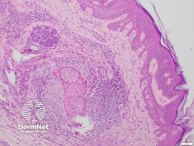
Epithelial sheath neuroma is diagnosed by histopathological examination of a skin biopsy. It is characterised by multiple enlarged peripheral nerve fibres that are sheathed by mature squamous epithelium. Epithelial sheath neuroma is sometimes surrounded by myxoid (mucus-like) stroma and a lymphocyticinfiltrate.
Histology of epithelial sheath neuroma
What is the differential diagnosis for epithelial sheath neuroma?
The clinical differential diagnosis for epithelial sheath neuroma may include:
The histological differential diagnosis for epithelial sheath neuroma includes reactive neuroepithelial aggregates, perineural invasion of a well-differentiated cutaneous carcinoma, or re-excision perineural invasion [7,8].
What is the treatment and outcome of epithelial sheath neuroma?
Epithelial sheath neuroma is benign.
The treatment of choice is excision, with no reports of recurrence after excision to date [1].
References
Flora A, Kim RH, Lara Rivero AD, Carr U, Isaacs F. Epithelial sheath neuroma: a case series. JAAD Case Rep. 2020;6(3):240-2. doi:10.1016/j.jdcr.2020.01.018. PubMed Central
Requena L, Grosshans E, Kutzner H, et al. Epithelial sheath neuroma: a new entity. Am J Surg Pathol. 2000;24(2):190-6. doi:10.1097/00000478-200002000-00004.PubMed
Zelger BG, Zelger B. Epithelial sheath neuroma: a benign neoplasm?. Am J Surg Pathol. 2001;25(5):696-8. doi:10.1097/00000478-200105000-00024. PubMed
Kutzner H. For Valentine's Day: epithelial sheath neuroma. Cancer. 2001;91(4):804-5. doi: 10.1002/1097-0142(20010215)91:4<804::Aid-cncr1067>3.0.Co;2-t. PubMed
Dunn M, Morgan MB, Beer TW, Chen KT, Acker SM. Histologic mimics of perineural invasion. J Cutan Pathol. 2009;36(9):937-42. doi:10.1111/j.1600-0560.2008.01197.x.PubMed
Wang JY, Nuovo G, Kline M, Magro CM. Reexcision perineural invasion and epithelial sheath neuroma possibly on a spectrum of postinjury reactive hyperplasia mediated by IL-6. Am J Dermatopathol. 2017;39(1):49-52. doi:10.1097/DAD.0000000000000671. PubMed
Chen KT. Reactive neuroepithelial aggregates of the skin. Int J Surg Pathol. 2003;11(3):205-10. doi:10.1177/106689690301100307. PubMed
Stern JB, Haupt HM. Reexcision perineural invasion. Not a sign of malignancy. Am J Surg Pathol. 1990;14(2):183-5. doi:10.1097/00000478-199002000-00010.PubMed