Epidermal naevus falls into the category of benignepidermaltumours.
Histology of epidermal naevus
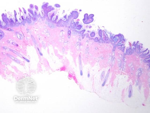
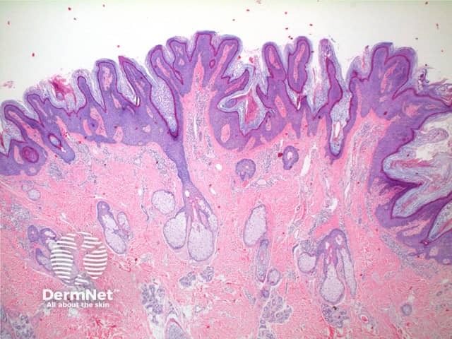
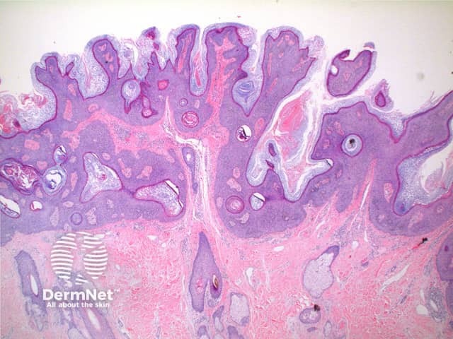
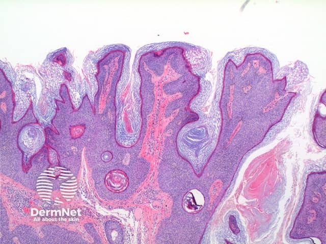
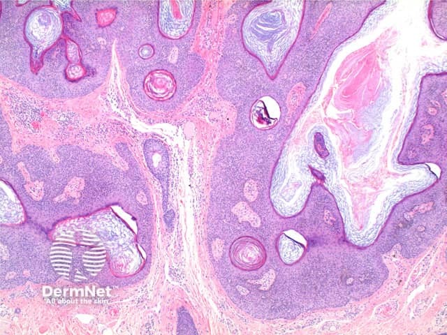
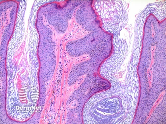
The scanning power view of an epidermal naevus is of an epidermal proliferative process (Figure 1). Low power reveals hyperkeratosis and papillomatosis across the breadth of the specimen (Figures 2,3). Epidermal hyperplasia forms finger-like projections with the intervening invaginations filled with hyperkeratotic material (Figure 4). Frequently incidental fungal spore forms are seen. Extending below the projections into the dermis are anastomosing thickened cords of the acanthotic epidermis (Figure 5). Dermalcollagen and telangiectatic vessels can be seen within the papillary projections (Figures 5 and 6).
A variable inflammatoryinfiltrates may accompany the epidermal changes, more prominent in the inflammatory verrucous variant.
Figure 1
Figure 2
Figure 3
Figure 4
Figure 5
Figure 6
Histological variants of epidermal naevus
A number of reported variants exist including acantholytic, porokeratotic, acanthosis nigricans-like, Hailey-Hailey disease-like, and verrucous epidermal naevus.
Differential diagnosis of epidermal naevus
Inflammatory Verrucous Epidermal Naevus (ILVEN) — this naevus is likely simply a subtype of the epidermal naevus. In addition to the superficial perivascular or lichenoidlymphocyticinfiltrate specific epidermal changes are recognised. Areas of alternating parakeratosis and orthokeratosis are seen. Beneath the parakeratosis there is hypogranulosis, whereas beneath the orthokeratosis there is hypergranulosis.
Seborrhoeickeratosis — while clinical correlation is essential here, the presence of elongated down-growths of epidermis with some flattening at the base is more in keeping with an epidermal nevus.
References
Skin Pathology (2nd edition, 2002). Weedon D
Pathology of the Skin (3rd edition, 2005). McKee PH, J. Calonje JE, Granter SR