
ADVERTISEMENT
You do not have any notes added to this page yet
Introduction Demographics Clinical features Diagnosis Treatment
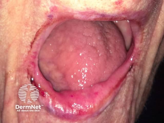
Eosinophilic ulcer of the oral mucosa is an uncommon benign ulcer seen in middle-aged to elderly adults that appears suddenly in the mouth or on the lips, is usually painful, and heals over a few weeks. It may represent a nonspecific reaction pattern to trauma.
Eosinophils are a type of inflammatory cell with a characteristic appearance under the microscope.
Eosinophilic ulcer of the oral mucosa is also known as traumatic ulcerative granuloma with stromal eosinophilia, oral traumatic granuloma, traumatic granuloma of the tongue or eosinophilic ulcer of the tongue. It is possibly the adult version of Riga-Fede disease.
Eosinophilic ulcer of the oral mucosa affects adults, with a peak in the sixth and seventh decades of life (50–80 years of age). There is a slight female predominance reported.
The cause of eosinophilic ulcer of the oral mucosa is unknown. Trauma is a reported trigger in 39%. The ulcer usually occurs on sites where trauma from teeth is common and it is seen in the age group most likely to have damaged teeth and dentures that may cause trauma. However, most simple traumatic ulcers in the mouth do not show the characteristic clinical and histological features of eosinophilic ulcer. Therefore it has been suggested that trauma may allow as-yet-unidentified infections, toxins or foreign proteins to enter and trigger the characteristic inflammatory reaction in susceptible people. Another theory is that it represents a CD30+ lymphoproliferative disorder (a type of lymphoma).
The typical clinical features of eosinophilic ulcer of the oral mucosa include:
The most commonly affected site is the tongue, usually the sides or the top.
Other reported locations (in decreasing order) are:
The patient is generally well, but eating and drinking may be limited by pain. Enlarged lymph glands in the neck may be felt in rare cases.
Eosinophilic ulcer of the oral mucosa usually heals by itself within one month but can persist up to one year. Recurrences have rarely been reported.
Atypical clinical presentations without an ulcer have included:
A biopsy is usually required to confirm this diagnosis and exclude other conditions, such as aphthous ulcers and most importantly oral cancer and infections.
The histology is characteristic:
Eosinophilic ulcer of the oral mucosa usually heals by itself within one month, so no specific treatment is required. Rapid healing is typically seen after a biopsy. Analgesics may be required for pain relief. Attention to rough teeth or dental prostheses may aid healing.
Reported treatments have included:
An AI summary will appear based on your search term using data from all of the topic pages across the entire DermNet site.
Show more