This distinctive tumour is an adnexal tumour argued to arise from the apocrine or eccrine secretory coil or coiled duct.
Histology of cylindroma
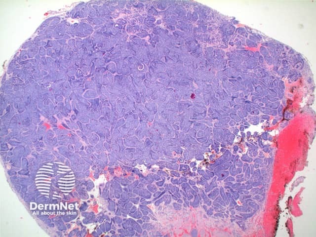
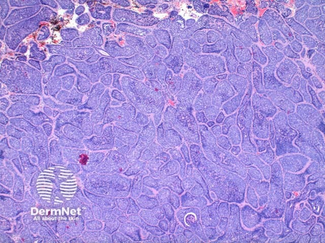
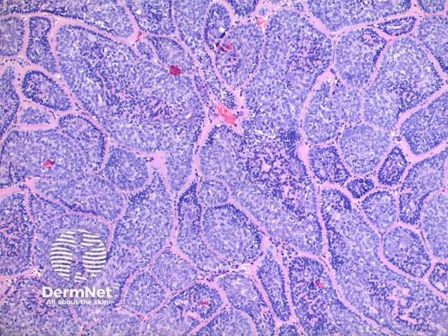
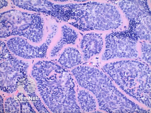
Low power view of cylindroma shows a non-encapsulated tumour nodule arising from the dermis (Figure 1). This is formed by multiple irregular tumour islands, distributed in an aptly named ‘jigsaw’ pattern (Figure 2). Surrounding the tumour islands, and in discrete droplets within the nodules is a thick hyaline deposit (Figure 3). Two populations of cells are noted to make up the tumour nodules. A smaller cell with a hyperchromaticnucleus tending to the periphery, and larger cells with open nuclei throughout the centre of the nodules (Figure 4).
Figure 1
Figure 2
Figure 3
Figure 4
Special stains for cylindroma
The hyaline bands and deposits are PAS positive and diastase resistant, and are known to contain type IV and type VII collagen.
Variants of cylindroma pathology
Brooke-Spiegler syndrome and multiple cylindromatosis turban tumour syndrome: No differences are seen between sporadic cylindromas or those occurring in the context of this syndromes. Tumours with overlap features with eccrine spiradenoma can be seen.
Differential diagnosis of cylindroma pathology
Eccrine spiradenoma: In this tumour the low power view shows a diffuseproliferation of the basophilic component while lacking the discrete tumour islands. There is frequently a minor lymphocyticinfiltrate present, while in cylindroma there is a more prominent population of S100 positive dendritic cells.
Spiradenocylindroma: In some tumours features of both cylindroma and eccrine spiradenoma can be seen and are designated as overlap tumours.
References
Skin Pathology (2nd edition, 2002). Weedon D
Pathology of the Skin (3rd edition, 2005). McKee PH, J. Calonje JE, Granter SR
Immunohistochemical differentiation of four benign eccrine tumors. Missall TA, Burkemper NM, Jensen SL, Hurley MY. J Cutan Pathol. 2009 Feb;36(2):190–6. PubMed
Expression of stem-cell markers (cytokeratin 15 and nestin) in primary adnexal neoplasms-clues to etiopathogenesis. Mahalingam M, Srivastava A, Hoang MP. Am J Dermatopathol. 2010 Dec;32(8):774–9. PubMed