Histology of combined trichoepithelioma and cellular blue naevus
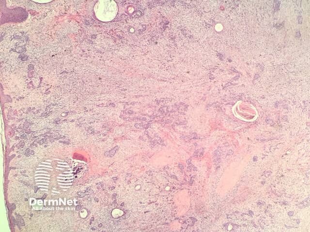
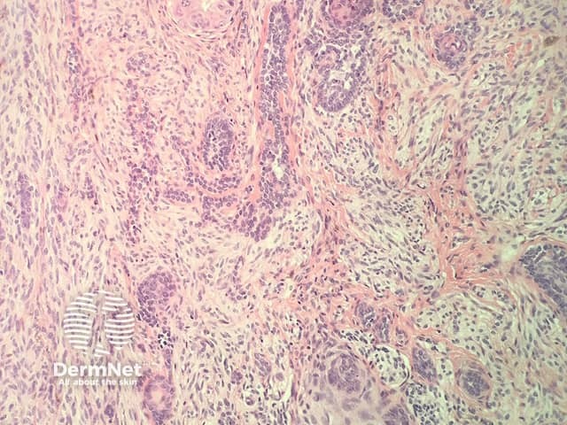
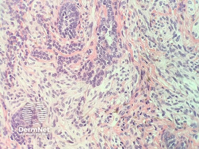
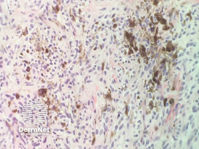
In combined trichoepithelioma and cellular blue naevus, the histopathology shows a fairly well-circumscribed biphasic dermalproliferation involving the full thickness of the dermis and shows tongues of rounded tumour fronts (figure 1). The tumour is composed of intimately mixed populations of melanocytes and basaloid cells. The basaloid cells anastomose in a lace-like pattern and form horncysts focally (figure 2,3). The follicular component is surrounded by a prominent stroma, which is condensed around the basaloid islands as hairpapilla-like structures. The melanocytic component consists of islands of plump spindled cells with abundant pale cytoplasm and minimal pigment and rarer slender dendritic melanocytes with heavy melaninpigmentation (figure 4).
Figure 1
Figure 2
Figure 3
Figure 4
Special studies for combined trichoepithelioma and cellular blue naevus
The follicular and melanocytic components stain strongly with p63 and S100, respectively, and there is no cross-over of staining.
The differential diagnosis for combined trichoepithelioma and cellular blue naevus
Other diagnoses to be considered include:
Malignant blue naevus — these usually show more evidence of nuclearatypia, proliferation and tumour necrosis
Malignant basomelanocytic tumour — these rare tumours show an intimate admixture of malignant basaloid cells and malignant melanocytes.
References
Martin R, Emanuel P. Combined cellular blue nevus and trichoepithelioma. J Clin Aesthet Dermatol. 2013 Aug;6(8):35–8. PubMed PMID: 24003350. PubMed.