Carcinoma erysipeloides is an uncommon form of cutaneous metastasis in which malignant cells spread to the skin via superficial dermallymphatic vessels.1
Carcinoma erysipeloides
Carcinoma erysipeloides
Carcinoma erysipeloides
Carcinoma erysipeloides
Carcinoma erysipeloides
Carcinoma erysipeloides
Carcinoma erysipeloides
Carcinoma erysipeloides
What are the clinical features of carcinoma erysipeloides?
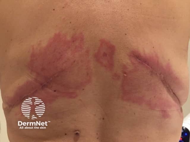
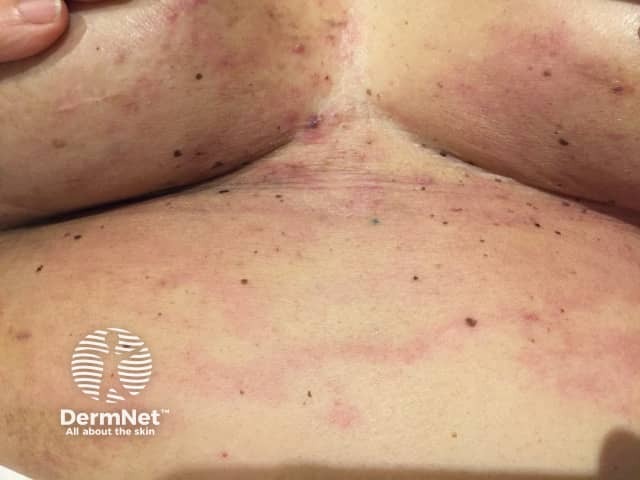
Carcinoma erysipeloides presents as a patch of thickened, red skin resembling cellulitis. Although usually tender, it can be asymptomatic and does not cause fever. Carcinoma erysipeloides is most commonly found on the chest. It may appear within a scar, involve an arm, or uncommonly, the head and neck region. In cases of breast cancer, the other breast may become involved. 2
The edge of a patch of carcinoma erysipeloides may be distinctly raised, red and swollen due to tumour cells blocking the lymphatic vessels and the release of cytokines.3
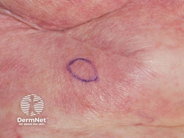
Carcinoma erysipeloides tends to occurs near the primary tumour. Signs that the cancer has spread to the skin include:
A firm, round or oval, mobile, non-painful nodule
Carcinoma en cuirasse, also called sclerodermoid carcinoma: indurated scar-like plaques due to cancer cells infiltrating collagen
Carcinoma telangiectoides: red patches with numerous blood vessels (telangiectases) or lymphatic vessels (lymphangiectasia) due to infiltration by cancer cells.
What causes carcinoma erysipeloides?
Most cases of carcinoma erysipeloides are due to underlying adenocarcinoma, most commonly adenocarcinoma of the breast.4 It has also been described with melanoma, and tumours of the parotid gland, thyroid, larynx, lung, fallopian tube, cervix, ovary, colon, prostate, pancreas and stomach.4-5 Rarely it can be the first sign of the tumour.4
Carcinoma erysipeloides usually appears after chemotherapy, radiotherapy or surgery to remove the tumour and/or local lymph nodes. It is thought that these treatments may lead to shedding of the tumor cells through the lymphatics.6
How do you diagnose carcinoma erysipeloides?
The diagnosis of carcinoma erysipeloides can be difficult. Delay in diagnosis is common, because it may resemble infection (cellulitis/erysipelas) or radiation dermatitis.7
It is not possible to remove carcinoma erysipeloides by surgery. Palliative treatment with chemo-radiotherapy may cause regression.9 The prognosis of a patient with carcinoma erysipeloides is generally poor.
References
Lever L, Holt P. Carcinoma erysipeloides. Br J Dermatol 1991; 124: 279–82
Gaffar BA, Almualla A. Post-mastectomy breast rash. International Journal of Dermatology 2010: 49; 855–857
Rook’s Textbook of Dermatology, 8th Edition, Edited by Tony Burns, Stephen Breathnach, Neil Cox, Christopher Griffiths, March 2010, Wiley-Blackwell
Nikolaou V, Stratigos A, Frangia K, Nikolaidis I, Syrigos K. Carcinoma erysipeloides deriving from a primary cutaneous squamous cell carcinoma . International Journal of Dermatology 2011, 50, 754–765
Cox SE, Cruz PD. A spectrum of inflammatory metastasis to skin via lymphatics: three cases of carcinoma erysipeloides. J Am Acad Dermatol 1994;30:304-7
Mackowiak PA. Rash in a Patient With Ovarian Cancer. Clinical Infectious Diseases 2012;54(4):538
Gugle A, Malpathak V, Zawar V, Deshmukh M, Kote R. Carcinoma erysipeloides: an unusual presentation mimicking radiation dermatitis. Dermatol Online J 2008;14:26
Nambi R, Tharakaram S. Carcinoma erysipeloides as a presenting feature of breast carcinoma. Int J Dermatol 1999;38:367-8
Finkel LJ, Griffiths CEM. Inflammatory breast carcinoma (carcinoma erysipeloides), an easily overlooked diagnosis. Br J Dermatol 1993;129:324–6.