
ADVERTISEMENT
You do not have any notes added to this page yet
Introduction Causes Demographics Clinical features Diagnosis Differential diagnoses Treatment
Calcaneal petechiae, also known as talon noir ('black heel'), are a benign condition due to the accumulation of blood in the stratum corneum of the heel. Many other terms have been used to describe these haemorrhagic lesions of the stratum corneum, including subcorneal haematoma, haemorrhagic hyperkeratosis, basketball heel, and tennis heel/toe. Similar lesions can be found on the hands, such as tache noir ('black palm') and PlayStation thumb.
Calcaneal petechiae follow repeated pounding or lateral shearing forces causing dilation and rupture of fragile blood vessels in the papillary dermis, micro-haemorrhages, and extravasation of blood cells through sweat ducts into the epidermis. Extravasated red blood cells in the stratum corneum are inaccessible to phagocytic cells. Jumping, sudden stopping, or twisting on the heel are the usual activities that precipitate the bleeding.
As several of the alternative names suggest, calcaneal petechiae are common in adolescent or young adult athletes. Many of the earliest cases were reported in basketball players.
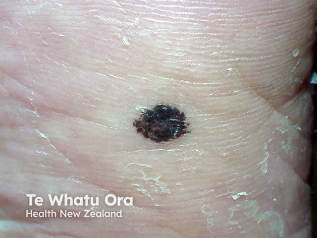
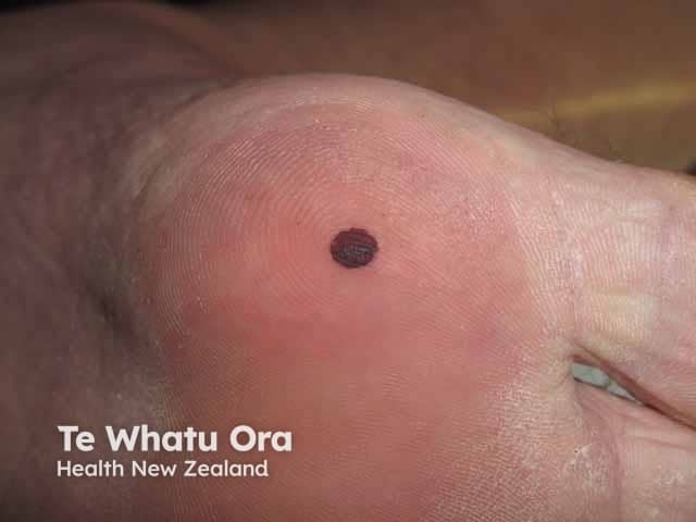
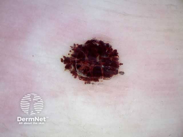
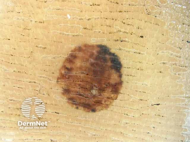
Calcaneal petechiae present as the sudden onset of lesions with intracorneal haemorrhage. These can vary in colour (red, brown, black), number, size (multiple pinpoint to large single lesions), and location (heel, sole, toe). Calcaneal petechiae are asymptomatic. Paring with a scalpel blade shows the colour is in the shavings and the colour is rapidly removed from the skin.
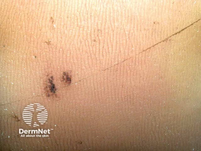
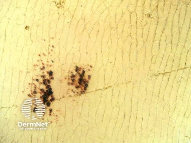
Dermoscopy of subcorneal haematomas demonstrates a red-brown colour; brown, red, and black are less commonly seen. The most common pattern of pigmentation is homogenous, followed by globular and parallel ridge. See also Dermoscopy of other non-melanocytic lesions.
Calcaneal petechiae are usually a clinical diagnosis supported by dermoscopy and paring of the lesions without need for a biopsy. Histological assessment of the lesion would show hyperkeratosis and epidermal acanthosis, with focal haemorrhage within the stratum corneum (see Talon noir pathology).
Appropriate diagnosis of calcaneal petechiae is important as the condition may mimic benign or malignant melanocytic lesions, including acral lentiginous melanoma. Other diagnoses to consider may include viral warts and traumatic tattoo.
Paring of the lesion helps distinguish calcaneal petechiae from these conditions:
Following diagnosis, calcaneal petechiae do not require treatment. Calcaneal petechiae resolve spontaneously within 4–6 weeks. They may recur if the triggering skin trauma continues. Paring of the stratum corneum with a scalpel blade will remove the haemorrhagic material without bleeding or pain.
An AI summary will appear based on your search term using data from all of the topic pages across the entire DermNet site.
Show more