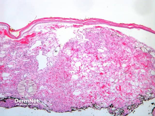
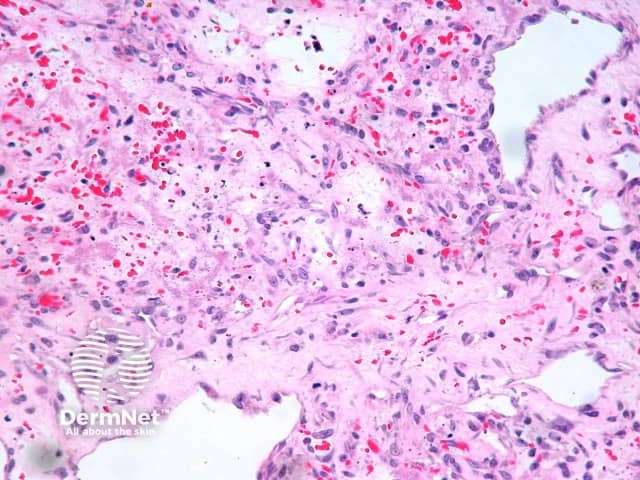
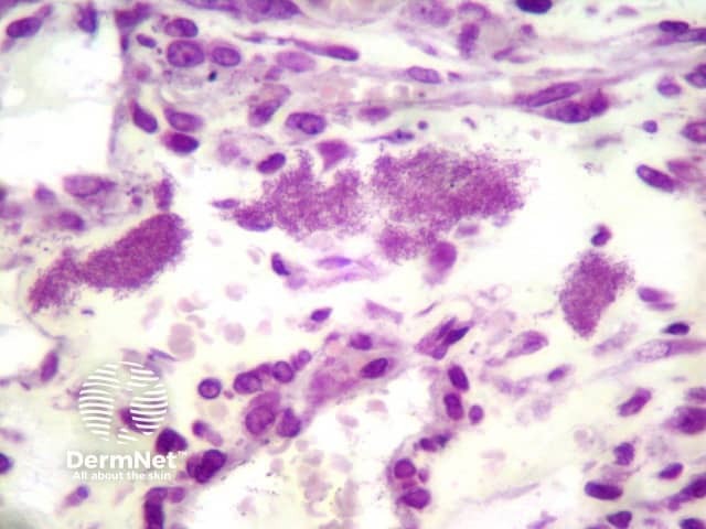
Histologically, the lesions of bacillary angiomatosis closely resemble pyogenic granuloma. Typically, there are ulcerated papules or nodules composed of lobules of highly vascularised oedematousconnective tissue (Figure 1). There is typically a mixed-cell infiltrate and nuclear dust of degenerated inflammatory cells. Accompanying this peculiar background are diagnostic clumps of pink-purplish granular material (Figure 2). This granular material consist of huge numbers of the gram-negative rods of Bartonella (Rochalimaea) henselae (Figure 3).
Figure 1
Figure 2
Figure 3
Special stains for bacillary angiomatosis
Warthin-Starry stain or gram stain highlight the pink-purple clumps of bartonella organisms. The organisms are revealed to be gram-negative rods with gram stain (Figure 3).
Differential diagnosis of bacillary angiomatosis pathology
Pyogenicgranuloma – This also shows an ulcerated lobulated vascularproliferation and inflammatory changes. The organisms are an obvious clue. In organism-poor cases fragmented inflammatory cells can be an important clue and prompt further microbiologic studies.
Verruga peruana – This disease is practically indistinguishable from bacillary angiomatosishistopathologically and similar clumps of bacteria may be found. This disease occurs in Peru and some other parts of South America and is caused by an infectious agent, Bartonella bacilliformis, a relative of Bartonella henselae.
References
DERM101.COM Ackerman, A. Bernard (2000)
Pathology of the Skin (Fourth edition, 2012). McKee PH, J. Calonje JE, Granter SR