Apocrine mixed tumour is also known as apocrine chondroid syringoma. These lesions present as non-descript dermalnodules which grow slowly. They most often arise on the skin of the head and neck.
Histology of apocrine mixed tumour
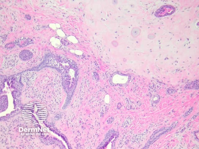
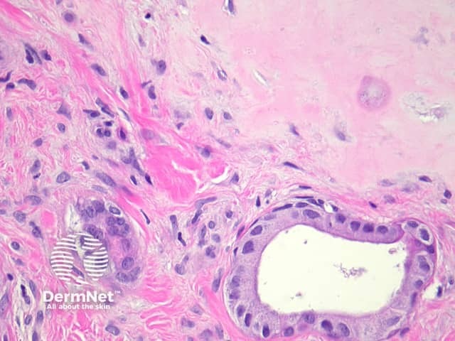
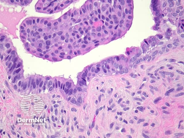
Apocrine mixed tumours form a well circumscribed dermal mass. These distinctive tumours are composed of both a mesenchymal component which is usually chondroid, and an epithelial component (figure 1). The cartilage is mature, with bland chondrocytes and a silver/grey matrix (figure 2). The epithelium is bland, and consists in most areas of two cell layers: a myoepithelial layer and a lining luminal layer (figure 3).
Figure 1
Figure 2
Figure 3
Special studies for apocrine mixed tumour
None are generally needed.
Differential diagnosis of apocrine mixed tumour
Malignant apocrine mixed tumour: Marked atypia, numerous mitoses and lymphovascular space invasion indicate malignant transformation (fortunately a rare event). Rare cases of bland appearing apocine mixed tumour have metastasized so some authorities advocate complete surgical excision of all tumours
Eccrine mixed tumour: These are composed of quite regularly spaced small glands lined by a thin epithelium set in a fibromucinous stromaPleomorphicadenoma: These salivarygland tumours are usually morphologically identical. The location in the parotid gland or minor salivary gland allows distinction
References
Constantinescu MB, Chan JB, Cassarino DS. Chondroid syringoma with tyrosine crystals: case report and review of the literature. Am J Dermatopathol. 2010 Apr;32(2):171–4. PubMed