
ADVERTISEMENT
You do not have any notes added to this page yet
Introduction Demographics Causes Clinical features Complications Diagnosis Differential diagnoses Treatment Outcome
A cutaneous angiofibroma is a benign vascular neoplasm composed of dermal fibrous tissue and blood vessels.
Angiofibroma is classified by association with a genetic disorder or according to its body site [1].
Angiofibromas are associated with the following genetic disorders:
Angiofibromas are more commonly acquired.
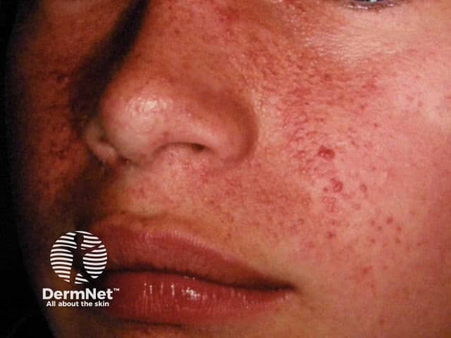
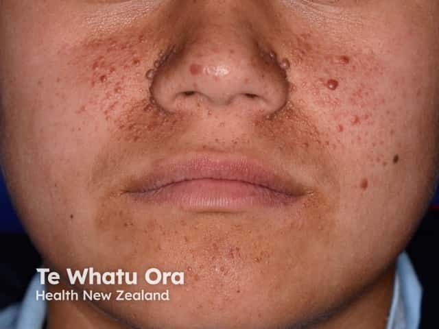
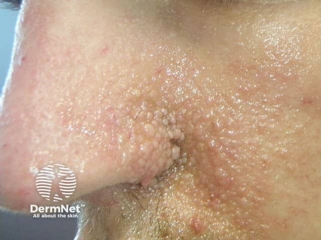
Tuberous sclerosis is a neurocutaneous autosomal dominant syndrome, in which angiofibromas appear in childhood in the nasolabial folds and on the central face [2]. Patients with tuberous sclerosis commonly develop an oral fibroma or a periungal angiofibroma (Koenen tumour) over time [1]. The facial angiofibromas associated with tuberous sclerosis are also called adenoma sebaceum, juvenile angiofibroma, and Pringle tumour.
Facial angiofibromas have been reported in Birt-Hogg-Dubé syndrome, a rare genodermatosis characterised by skin and renal tumours, as well as spontaneous pneumothorax [3]. Most of the cutaneous lesions however are fibrofolliculomas, which are abnormal growths of the hair follicles.
Multiple endocrine neoplasia type 1 is a hereditary syndrome that leads to tumours in several endocrine organs [1].
Angiofibromas can also be acquired and unrelated to a genetic syndrome, commonly in the form of:
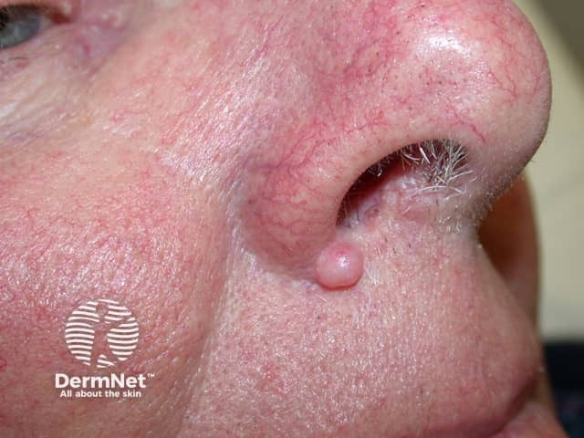
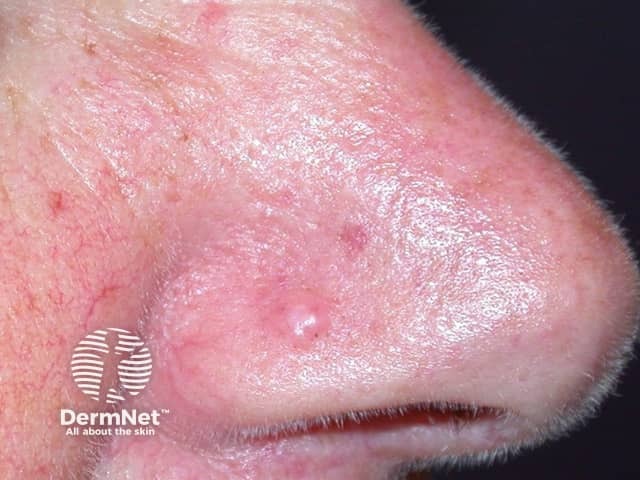
A fibrous papule is characteristically found in adults as a solitary lesion usually on the nose, often clinically mistaken for a basal cell carcinoma or melanocytic naevus. It is thought to be a form of dermal naevus.
Multiple pearly penile papules occur in 10–30% of adult males on the coronal edge and sulcus. They can be mistaken for viral warts [1,2].
See penile pearly papules images.
Angiofibromas are caused by a local overgrowth of collagen, fibroblasts, and blood vessels.
Genetic mosaicism for these genetic conditions must also be considered [4]. What specifically triggers the development of angiofibroma is unknown.
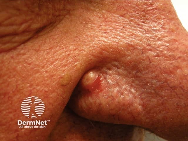
An angiofibroma is a firm, flesh-coloured dome-shaped papule less than 5 mm in diameter. Small capillaries may be visible on the surface of the lesion.
Angiofibromas may be itchy and may also bleed. Those associated with genetic syndromes result in facial disfigurement and stigmatisation [1]. See Psychosocial factors in dermatology.
The diagnosis of angiofibroma may be made clinically or after a skin biopsy. The histopathology of angiofibroma shows an ‘onion skin’ pattern around vessels and follicles, hyperkeratosis, and vascular proliferation [5].
If an underlying genetic condition is suspected, appropriate genetic screening and evaluation are required [1].
The differential diagnosis for angiofibroma depends on its location [1].
Differential diagnoses for facial lesions that can resemble angiofibromas can include:
Differential diagnoses for periungual lesions that can resemble angiofibroma can include:
Differential diagnoses for penile lesions that can resemble angiofibroma can include:
Angiofibromas are benign and do not always require removal. Options for treatment of angiofibromas include:
Multiple treatments are often necessary [1].
Although angiofibromas are benign, they are persistent. Angiofibromas can be removed for cosmetic or pain-related reasons. The recurrence rate for angiofibromas associated with tuberous sclerosis may be as high as 80% [1].
An AI summary will appear based on your search term using data from all of the topic pages across the entire DermNet site.
Show more