Author: Naomi Ashman, Dermoscopist, Torbay Skin, Auckland, New Zealand; DermNet New Zealand Editor in Chief: Adjunct A/Prof Amanda Oakley, Dermatologist, Hamilton, New Zealand. Copy edited by Gus Mitchell. May 2019.
Amelanoticmelanoma is a form of melanoma in which the malignant cells produce little to no pigment.
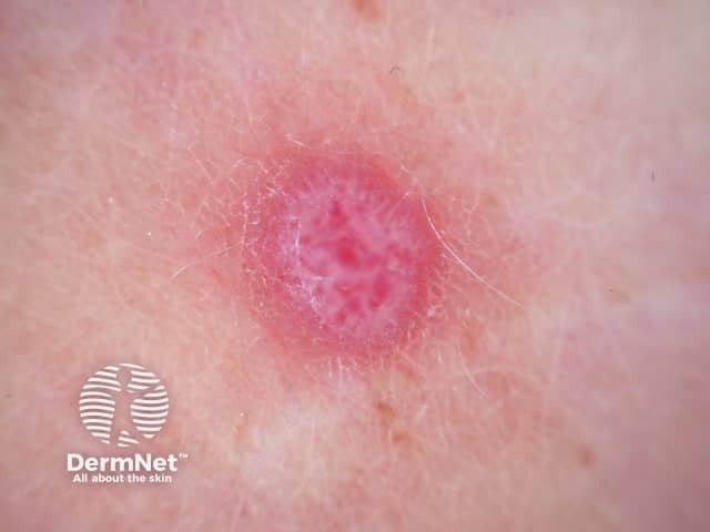
What are the clinical features of amelanotic melanoma?
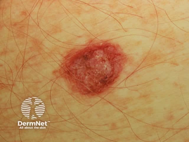
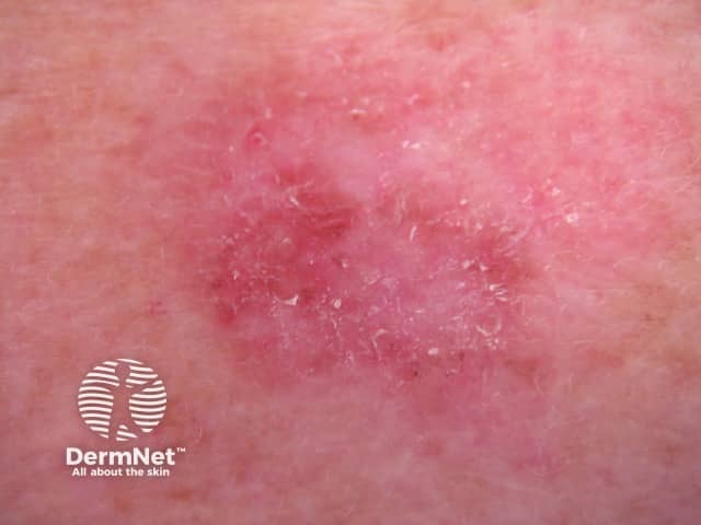
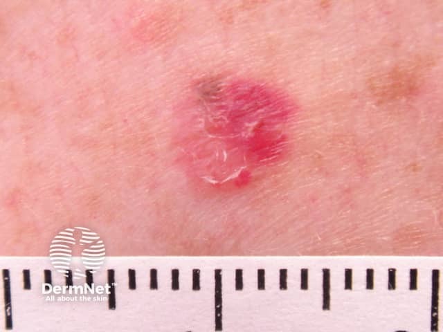
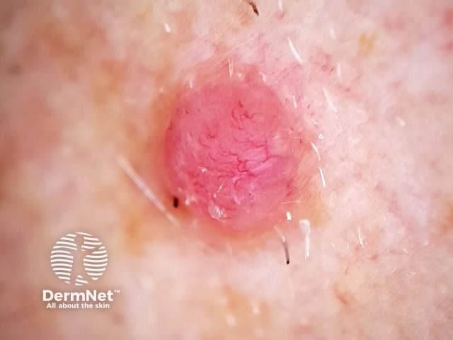
Amelanotic melanomas are classically described as skin coloured. A significant proportion of melanomas are red or pink. Typical early lesions present as asymmetricalmacules that may be uniformly pink or red and may have a faint light tan, brown, or grey pigmentation at the periphery. The borders may be well or ill-defined. Use of the 3 R’s (Red, Raised, Recent change), may help screen for amelanotic melanoma.
Amelanotic melanoma
Amelanotic melanoma
Amelanotic melanoma
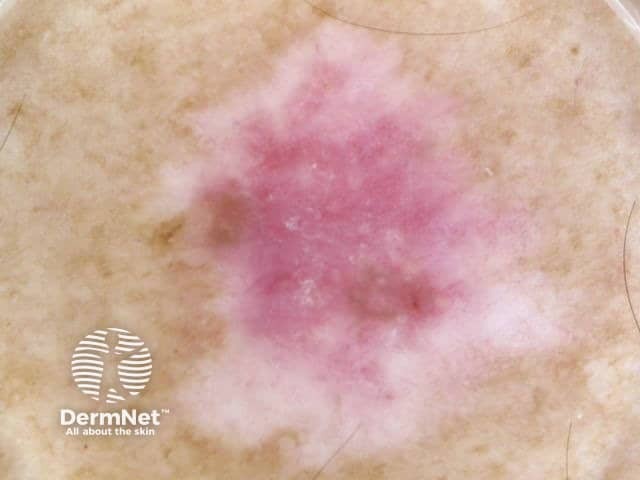
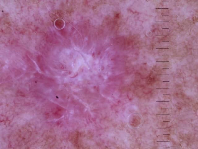
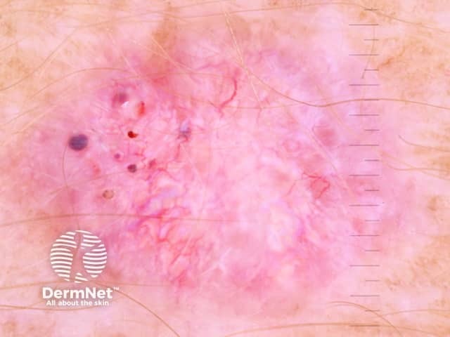
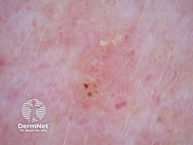
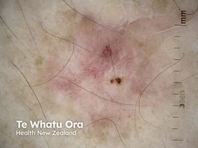
What are the dermoscopic features of amelanotic melanoma?
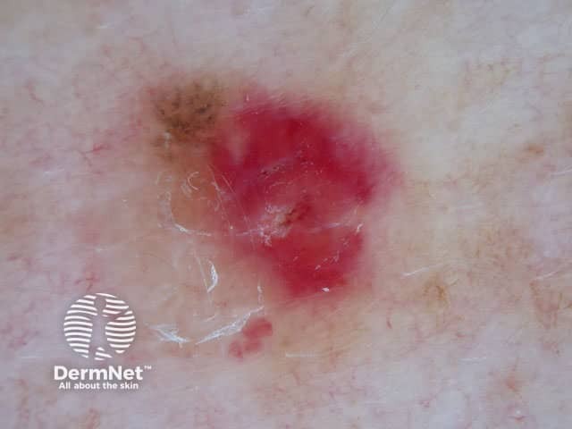
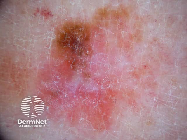
The dermoscopic features of amelanotic melanoma may include:
Irregular pigmentation/pigment remnants (if any is present)
Irregular dots or globules (in pigmented areas)
Polymorphousvascular pattern, especially if irregular dot vessels are present, or a combination of dotted and linear irregular vessels (the most frequent dermoscopic finding); helical vessels are strongly specific for melanoma
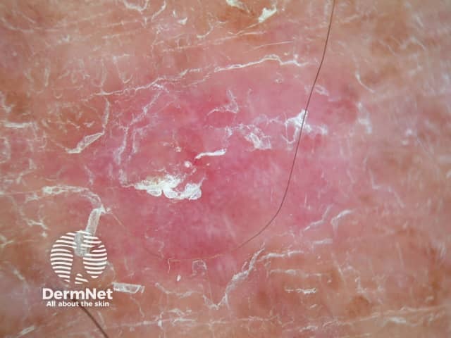
Multiple shades of pink
Milky red areas or clods
Reticulardepigmentation
White structureless areas
White lines on polarised dermoscopy.
Vessel analysis using the 5 + 2 List [1].
Vessel morphology should be evaluated carefully if lesions are lacking pigmentation. In the 5 + 2 list below, the presence of a certain type of vessel is asked for; if the answer is “no”, proceed to the next question.
If question number 5 is answered in the affirmative (ie, point-like or loop-like vessels are present), evaluate the absence of white halos and presence of traces of melanin. If “yes,” suspicion of melanoma is raised. Traces of brownish melanin, which may not be visible clinically, present focally within the lesion support the diagnosis of melanoma or Spitz nevus. It can be very difficult to discriminate between amelanotic melanoma and Spitz naevus by dermoscopy. Lesions with point-like or loop-like vessels without white halo must be considered suspicious and removed [1].
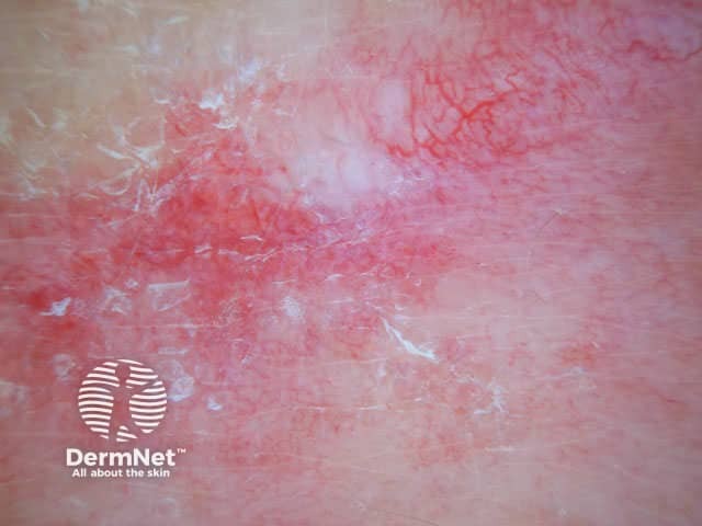
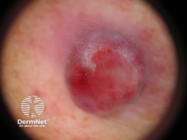
Multiple shades of pink
Linear irregular vessels
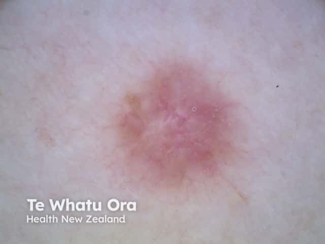
Pigment remnants
Milky pink areas
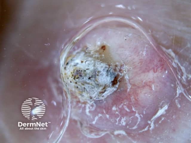
White structureless area, linear irregular vessels
White lines, polymorphous vessels
What is the differential diagnosis of amelanotic melanoma?
What is the histological explanation of amelanotic melanoma?
Histopathologically, an amelanotic melanoma is usually composed of highly malignant epithelioid cells. These cells display pleomorphism, mitotic activity, enlargement, large nucleoli, and lack of maturation as the cells descend into the dermis.
References
Kreusch J, Koch F. [Incident light microscopic characterization of vascular patterns in skin tumors]. Hautarzt 1996; 47: 264–72. Review. German. Erratum in: Hautarzt 1996; 47: 540. PubMed