A naevus (American spelling nevus, nevi) is a circumscribed and stable malformation of a component of the skin. Naevi (American spelling nevi) are often present at birth, when they are often called brown birthmarks. Naevi composed of melanocytes (the pigment cells that produce melanin) are called melanocytic naevi or pigmented naevi.
What are Ota, Ito and Hori naevi?
Naevus of Ota, naevus of Ito and naevus of Hori are special melanocytic naevi that have a slate-brown or blue/grey colouring. They are forms of dermal melanocytosis in which naevus cells are found deep within the dermis.
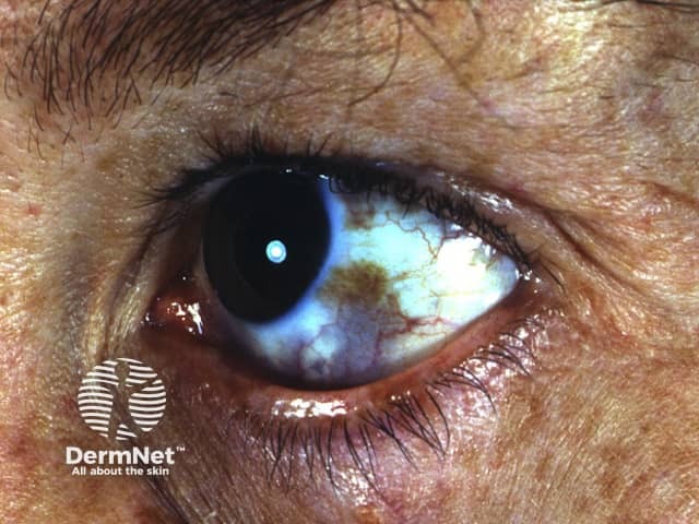
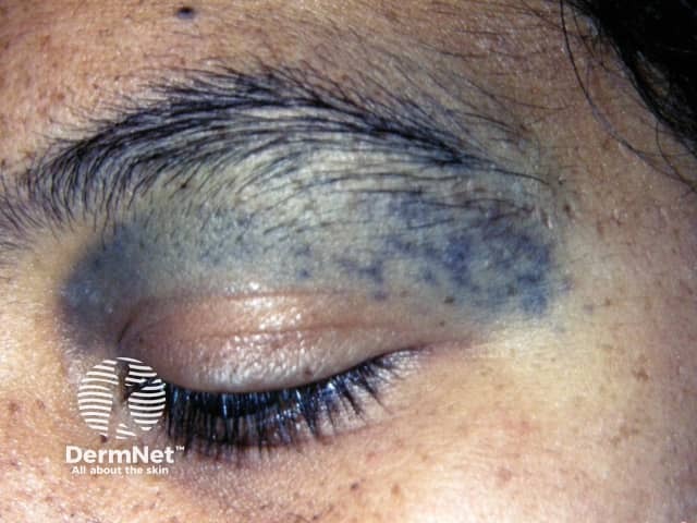
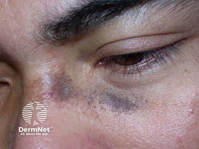
Naevus of Ota is on the forehead and face around the eye area. Hyperpigmentation of parts of the eye may occur (sclera, cornea, iris, retina)
Naevus of Hori is similar to naevus of Ota but affects both sides of the face
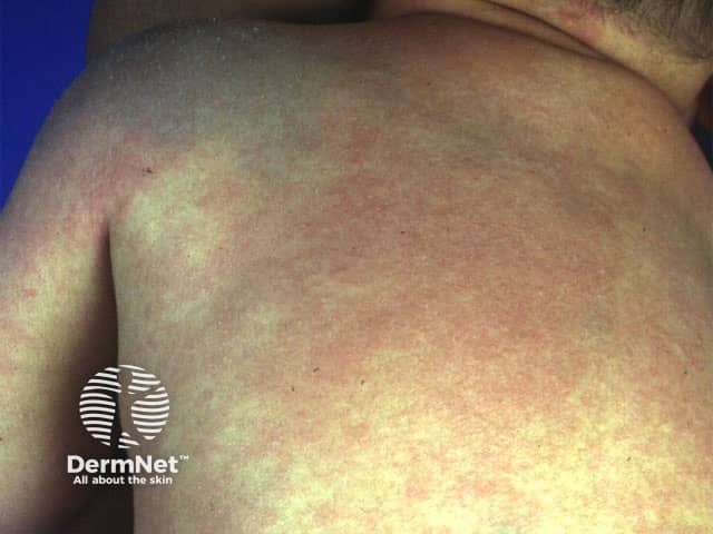
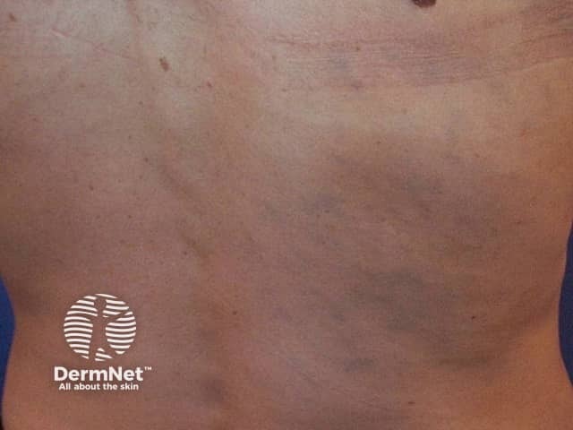
Naevus of Ito is on the shoulder and upper arm area (shoulder girdle).
Dermal melanocytosis can also occur elsewhere on the body, including inside the mouth.
Naevus of Ota
Naevus of Ota
Naevus of Ota
Naevus of Ota
Naevus of Ito
Naevus of Ito
Naevus of Ito
How does dermal melanocytosis arise, and who is at risk?
It is not known why dermal melanocytosis occurs. Specific mutations have been detected within the dermal melanocytes, most often GNAQ or GNA11. Researchers have suggested that hormones play a part in their development. The role of ultraviolet radiation is thought to be small, as it does not reach deep dermal melanocytes.
Naevus of Ota is much more common than naevus of Ito. These naevi are present at birth in 50% of cases but may appear during adolescence or adult life. Naevus of Hori is not present at birth and is therefore a form of acquired melanocytosis.
Naevi of Ota and Ito are most commonly found in Asian populations; 0.2–0.6% of Japanese people have a naevus of Ota. They appear more frequently in females. Both forms of naevi are uncommon in Caucasians.
What are the signs, symptoms and complications of dermal melanocytosis?
In all forms of dermal melanocytosis:
Colour may vary to include brown-violet, violet-blue or blue-green hues
Naevi present in childhood may slowly grow and darken until adulthood is reached
Colour or perceived colour of naevi may change according to personal and environmental conditions, e.g. fatigue, menstruation, hot weather
If affecting the eye, melanocytosis rarely causes glaucoma
Melanoma very rarely develops within dermal melanocytosis, and has usually been reported in Caucasians. Ocular melanoma has rarely been reported in the choroid, brain, orbit, iris, ciliary body, and optic nerve in association with a nevus of Ota.
How is the diagnosis of dermal melanocytosis made?
The diagnosis of dermal melanocytosis is usually made by observing typical discolouration of the skin. It is classified according to the site affected.
Some patients may undergo skin biopsy, which confirms the presence of melanocytes in the dermis.
Other skin conditions resulting in bluish or grey coloured skin may be considered. These include: