
ADVERTISEMENT
You do not have any notes added to this page yet
Introduction
Demographics
Causes
Clinical features
Complications
Diagnosis
Differential diagnoses
Treatment
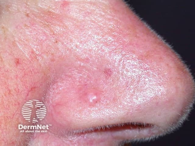
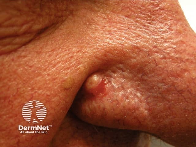
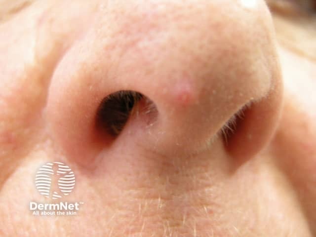
A fibrous papule of the nose is a common and harmless skin lesion. It is a firm solitary papule that occurs on or around the nose and has a characteristic appearance under the microscope.
A fibrous papule of the nose may also be known as a fibrous papule of the face, solitary angiofibroma, or sporadic angiofibroma.
Fibrous papules are common and may affect anyone. They usually appear during late adolescence and early adulthood. Multiple fibrous papules can be present without any underlying disorder.
The presence of multiple fibrous papules may be associated with genetic conditions including tuberous sclerosis, Birt-Hogg-Dubé syndrome, and multiple neuroendocrine neoplasia type 1 (MEN1). In these conditions, the papules are numerous and widespread, often beyond the nasal area. They are harmless apart from the psychological problems they may cause due to their visible appearance.
The cause of fibrous papules is not known.
They are often a single solitary lesion on or around the nose. The fibrous papule of the nose is dome-shaped, firm, non-tender, measuring approximately 1–6 mm in diameter. It can be skin-coloured, pigmented, white, or red in colour.
Fibrous papules are benign with no malignant potential.
A fibrous papule is diagnosed by skin biopsy.
There are distinctive microscopic features including:
Many of the histological features may also be seen in other types of angiofibromas and it can be difficult to distinguish them histologically. For this reason, a fibrous papule is currently considered to be a variant of angiofibroma. There are several histological subtypes of fibrous papules including:
See also the clear cell fibrous papule pathology page.
Fibrous papules may be similar in appearance to other skin lesions. They can be differentiated via biopsy and other clinical features:
Fibrous papules are harmless, do not require any treatment, and remain stable. They may be removed for cosmetic reasons by shave biopsy, excision, cryotherapy, laser therapy, and electrosurgery.
Once removed, fibrous papules rarely recur.
An AI summary will appear based on your search term using data from all of the topic pages across the entire DermNet site.
Show more