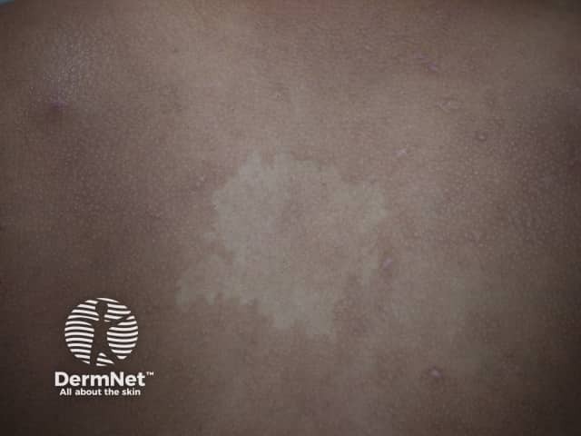
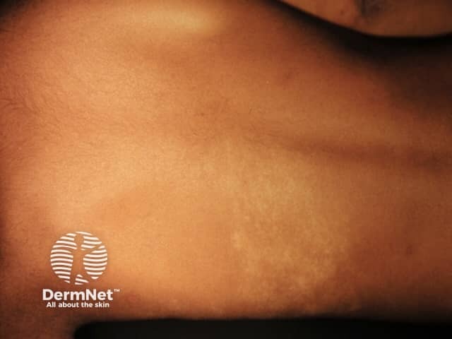
Achromic naevus is an uncommon birthmark (naevus) characterised by a well-defined pale patch. This is usually several centimetres in diameter, with an irregular but well-defined border. Shape and size varies. Often, smaller hypopigmentedmacules arise around the edges, resembling a splash of paint.
Achromic naevus (American spelling nevus) is also called naevus depigmentosus and non-pigmented naevus. The name is not quite right, as the hypomelanotic patches of an achromic naevus are not completely white, unlike the areas of depigmentation in vitiligo, which are amelanotic, and completely lacking melanocytes. Achromic naevi are usually solitary, in contrast to tuberous sclerosis, where multiple pale patches occur and are called ash-leaf spots.
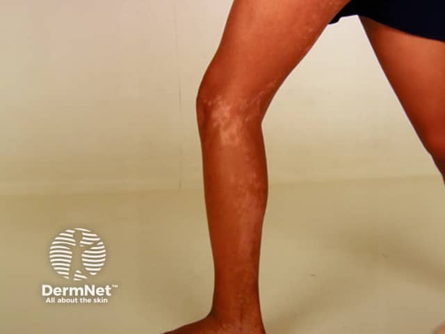
Achromic naevus is usually noted at birth or early childhood, although lesions may not be apparent until mid-childhood in those with light-coloured skin. The naevus remains stable over time apart from growing with the child. Achromic naevus most commonly arises on the trunk, but may also arise on the limbs and elsewhere. It is solitary in 50% of cases and may follow Blaschko lines.
Achromic naevus
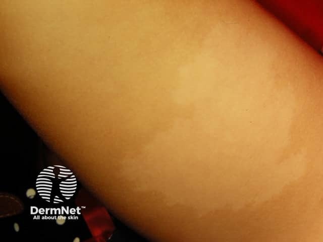
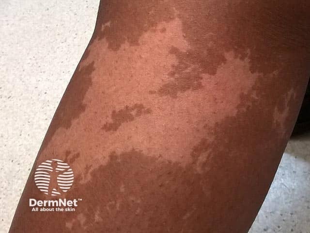
A unilateral depigmented lesion on the arm - the child was otherwise well. Now termed segmentalpigmentation disorder
What is the cause of achromic naevus?
Achromic naevus is a form of cutaneous mosaicism. It is caused by an altered clone of melanocytes (pigment cells) with a decreased ability to make melanin (brown pigment). Melanocyte numbers are normal or reduced in number, Melanosomes may be reduced in the melanocytes and/or keratinocytes suggesting impaired transfer.
What are the variants of achromic naevus?
Variants of achromic naevus include:
Isolated achromic naevus
Segmental achromic naevus, also called segmental depigmentation disorder, has midlinedemarcation and poorly-defined lateral borders; it is considered the hypopigmented form of “segmental pigmentation disorder”
Linear or ‘systematised’, achromic naevus has cutaneous findings that overlap with hypomelanosis of Ito.
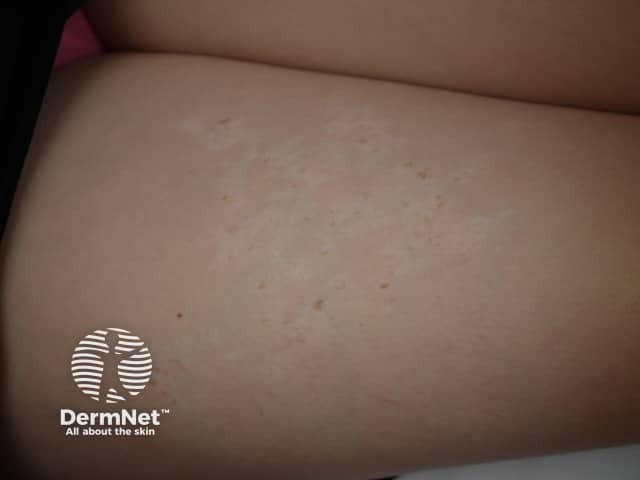
Occasionally, achromic naevus is associated with other neurocutaneous disorders. Co-localisedlentigines have been reported and are seen in figure 3 (top right) above. They may represent a reverse mutation. The ash-leaf macules seen in tuberous sclerosis are oval-shaped hypopigmented macules and look similar to achromic naevi but usually present as multiple lesions.
How is achromic naevus diagnosed?
Coupe identified diagnostic criteria for achromic naevus in 1976:
The patch of pale skin is present at birth or early in life
It remains in the same site throughout lifetime
There is no alteration in texture or change in sensation in the lesions
There is no dark border around the affected skin
Wood lamp examination: achromic naevus appears off-white, compared to the chalk-white accentuation seen in vitiligo.
Bolognia Dermatology 3rd Edition — Vitiligo and other disorders of hypopigmentation.
Kim SK, Kang HY, Lee ES, Kim YC. Clinical and histopathologic characteristics of nevus depigmentosus. J Am Acad Dermatol. 2006;55(3):423–8. doi:10.1016/j.jaad.2006.04.053. Medline.
Textbook of Pediatric Dermatology 2nd edition Harper J, Oranje A, Prose N. Blackwell Publishing 2006
Coupe RL. Unilateral systematized achromic naevus. Dermatologica 1967;134(1):19–35. doi:10.1159/000254235. PubMed
Lee HS, Chun YS, Hann SK. Nevus depigmentosus: clinical features and histopathologic characteristics in 67 patients. J Am Acad Dermatol 1999;40(1):21-6. doi:10.1016/s0190-9622(99)70524-4. Medline.
Mulekar SV, Al Issa A, Al Eisa A. Nevus depigmentosus treated by melanocyte-keratinocyte transplantation. J Cutan Aesthet Surg. 2011;4(1):29–32. doi:10.4103/0974-2077.79185. Medline.
Jagia R, Mendiratt V, Koranne RV, Sardana K, Bhushan P, Solanki RS. Colocalized nevus depigmentosus and lentigines with underlying breast hypoplasia: a case of reverse mutation?. Dermatol Online J. 2004;10(1):12. PubMed