
ADVERTISEMENT
You do not have any notes added to this page yet
Introduction Common sites Demographics Causes Signs and symptoms Tests Staging Treatment Prognosis
Melanoma develops if there is uncontrolled growth of melanocytes, the cells responsible for pigmentation. The majority of melanomas are cutaneous, ie they grow within the skin.
Mucosal melanoma is a rare type of melanoma that occurs on mucosal surfaces. Mucous membranes are moist surfaces that line cavities within the body. This means that mucosal melanoma can be found in the respiratory tract, gastrointestinal tract or genitourinary tract.
Mucosal melanomas are most often found in the head and neck, in the eyes, mouth, nasopharynx and larynx, but they can also arise throughout the gastrointestinal tract, anus and vagina. They make up approximately 1.1% of all melanomas but are usually more complicated because of late diagnosis due to their less visible locations and because they are often amelanotic – meaning they are not pigmented.
Subtypes of mucosal melanoma are based on the tissue in which they arise. Melanoma can start within any mucosal epithelium. The most common sites are:
The peak age of diagnosis of mucosal melanoma is between 70 and 79. However, younger people have also been known to develop mucosal melanoma, especially of the oral cavity.
Mucosal melanoma of the genital tract is more common in females.
While there are many suggested risk factors for mucosal melanoma, there is only weak evidence for all, and none that are widely accepted. About 25% of mucosal melanomas have been linked with problems with a gene called KIT. Genes are the templates used for making protein, and mutations in the KIT gene cause the production of a mutant protein. The KIT gene can also be over-expressed, which means that there is more of the KIT protein being made than usual. Both the mutation and over-expression of the KIT gene have been associated with mucosal melanoma.
Possible risk factors include:
The signs and symptoms of mucosal melanoma largely depend on its location. Therefore, there are a wide variety of symptoms that patients may experience.
If there is suspicion of mucosal melanoma, the most important test is a biopsy of the involved tissue. If the pathology report indicates melanoma, the patient will be fully examined and further tests may be undertaken to find out if it has spread further. These may include:
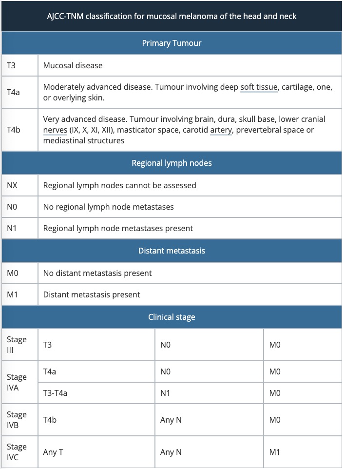
The American Joint Committee on Cancer (AJCC) Tumour, Node, and Metastasis (TNM) classification is used to stage mucosal melanoma of the head and neck. Mucosal tumour staging begins at T3 (advanced). Like cutaneous melanoma, mucosal melanoma can also spread to lymph nodes or through the blood stream to other areas of the body. Here, they can form new tumours. These are referred to as metastases.
Vulval melanomas are staged using the same AJCC-TNM classification as the table above. There is no staging system for vaginal melanoma, so a simplified clinical staging system can be used to categorise these melanomas.
Stage I |
Clinically localised disease |
||
Stage II |
Regional nodal involvement |
||
Stage III |
Distant metastatic involvement |
||
Anorectal mucosal melanoma is staged using the same simplified staging system as vaginal melanoma.
Stage I |
Clinically localised disease |
||
Stage II |
Regional nodal involvement |
||
Stage III |
Distant metastatic involvement |
||
The best form of treatment for mucosal melanoma is wide local excision of the lesion. This may not always be possible, as the melanoma may be located on an important anatomical structure, or be too large to excise safely.
Due to the likelihood of the same melanoma recurring, surgical resection is often combined with radiotherapy. Radiotherapy is also a consideration for patients who are not suitable for surgery.
Mucosal melanomas have a poor prognosis, as most patients develop metastases despite aggressive therapy.
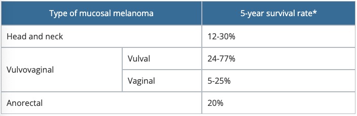
*The 5-year survival rate refers to the percentage of patients that survive for at least 5 years after their diagnosis, and is approximate.
An AI summary will appear based on your search term using data from all of the topic pages across the entire DermNet site.
Show more