
ADVERTISEMENT
You do not have any notes added to this page yet
Introduction
Demographics
Causes
Clinical features
Complications
Diagnosis
Differential diagnoses
Treatment
Outcome
Zygomycosis is any infection caused by fungi in the class Zygomycetes commonly found in soil or decomposing vegetation and animal matter. There are two major orders of Zygomycetes that cause infection in humans — Mucorales and Entomophthorales.
Zygomycosis due to Mucorales is seen predominantly in immunocompromised individuals. Diabetes mellitus is the leading predisposing cause worldwide, followed by haematological malignancies and stem cell transplants. Other associations include immune compromise due to solid organ cancer or transplant, drug-induced immunosuppression, or HIV (rare), and iron overload. In up to 20% of cases, the patient is immunocompetent and infection may complicate impairment of the skin barrier such as following skin trauma, thermal burns, or toxic epidermal necrolysis, and has been reported in premature infants. Graft-transmission of infection is an important route of acquisition in some countries.
The annual incidence ranges from approximately 1/million in the US and Europe to 140/million in India.
COVID-19-associated infection with Mucorales (‘black fungus’) has particularly affected diabetic males treated with systemic corticosteroids in India during the second wave of COVID-19 (April to July 2021), resulting in over 45000 cases and at least 4000 deaths.
Entomophthorales skin infection occurs in healthy individuals particularly around the tropical and subtropical humid areas such as in equatorial Africa and Southeast Asia.
The order Mucorales is divided into many families of which the two major causes of infection are Mucoraceae and Cunninghamellaceae. Within these families there are three important genera that cause infection in humans — Rhizopus, Mucor, and Lichtheimia (formerly Absidia) account for 75% of infections. Mucormycosis is the opportunistic infection caused by members of the Mucoraceae family.
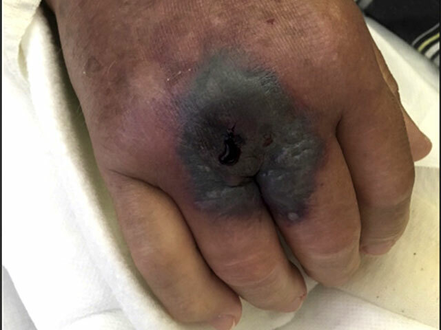
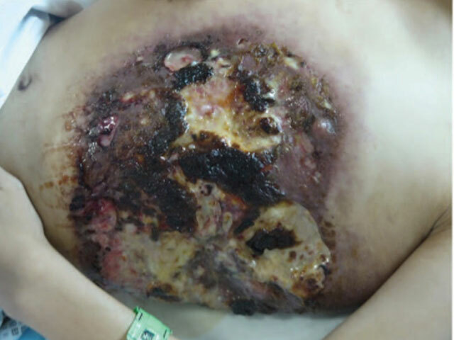
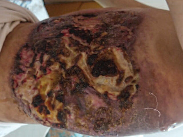
Primary cutaneous infection generally occurs due to direct implantation of fungal elements following major or minor skin trauma, such as skin abrasion, or tattoo, or in hospital following surgery, IV-line placement, or contaminated wound dressing. Secondary cutaneous involvement follows haematogenous dissemination.
Entomophthorales are insect pathogens. Two genera of the order Entomophthorales cause subcutaneous phycomycosis in humans, Basidiobolus and Conidiobolus.
There are five major clinical forms of mucormycosis:
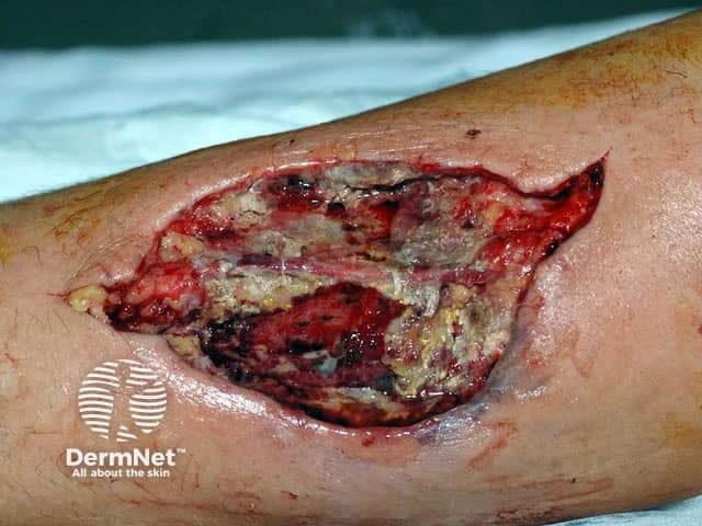
Cutaneous mucormycosis may be primary at the site of inoculation, or secondary in disseminated mucormycosis. Onset may be insidious or fulminant — acute and rapid in nature leading to gangrene.
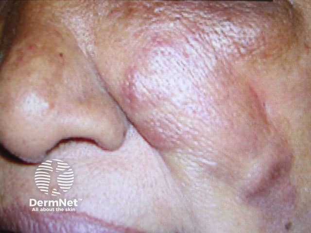
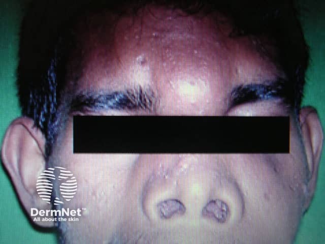
'Red flag' warning signs for rhinocerebral mucormycosis in an at-risk patient include:
Conidiobolus coronatus/incongruous |
|
Basidiobolus ranarum |
|
Diagnosis of cutaneous zygomycosis may be suspected clinically in an at-risk patient and confirmed on deep incisional skin biopsy with sufficient tissue for histology and culture.
Wet mount direct microscopy is a quick and inexpensive diagnostic test for mucormycosis on samples from usually sterile body tissues.
Zygomycosis can be difficult to treat despite the development of new antifungal agents. Treatment should be started within 6 days of symptom-onset for the most favourable outcome. Mucormycosis is often fatal although primary cutaneous disease has a lower mortality rate than gastrointestinal or pulmonary infection. In the past mortality rates were as high as 80% and even now the 90-day mortality rate is around 40%. Disseminated mucormycosis still has a mortality rate of over 90%. Rhinocerebral zygomycosis in solid organ transplant recipients is almost always fatal due to the high rate of CNS invasion.
Spontaneous resolution of entomophthormycosis has been reported. Recurrence is common even after surgical excision. The course is typically indolent.
An AI summary will appear based on your search term using data from all of the topic pages across the entire DermNet site.
Show more