Piebaldism is a rare inherited disorder of pigmentation characterised by patchy leukoderma (white skin) and white hair (poliosis) present at birth.
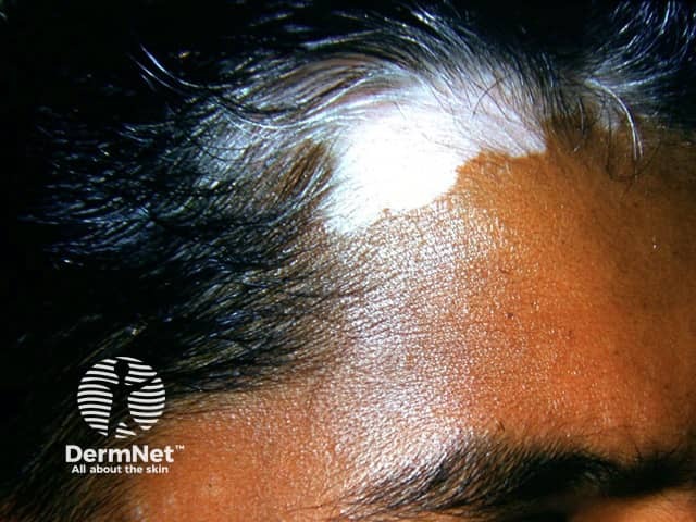
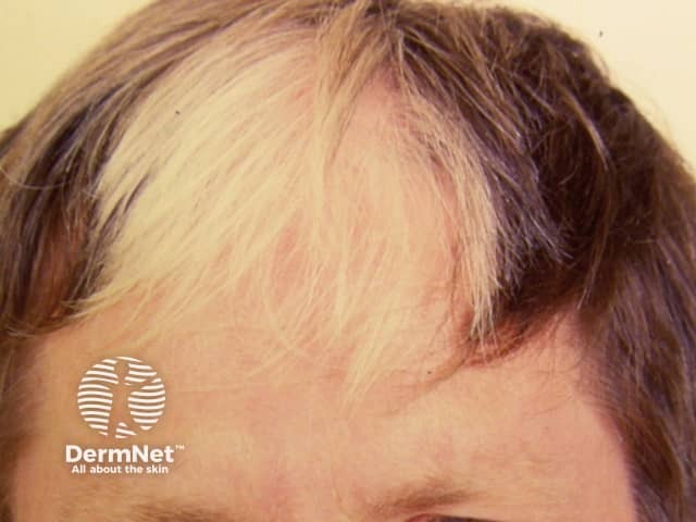
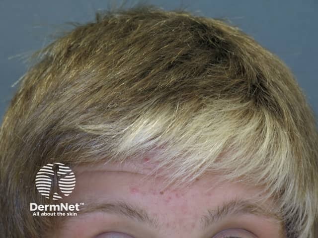
The name piebaldism is derived from a combination of the “pie” as in the magpie (a bird of black and white plumage) and the “bald” of the bald eagle (the US national bird that has a white feathered head). Hence the major characteristic of piebaldism is a white forelock (a patch of white hair directly above the forehead).
Who gets piebaldism?
Piebaldism is an autosomal dominant genetic condition, so each child of an affected parent has a 50% chance of inheriting the disorder.
What is the cause of piebaldism?
Piebaldism is a neurocristopathy caused by mutations of the KIT proto-oncogene on chromosome 4 account for75% of cases; over 45 different point mutations, deletions, nucleotide splice mutations, and insertions of the KITgene have been identified. The KIT gene mutation causes aberrant migration of melanoblasts from the neural crest to the skin in the embryo resulting in patches of skin lacking melanocytes. The severity of the condition correlates with the specific mutation within the KIT gene.
What are the clinical features of piebaldism?
White forelock in 80-90% of those affected (poliosis)
Leukoderma (white patch due to absence of melanocytes) of the central portion of the forehead
Eyebrow and eyelash hair may also be affected, either continuously or discontinuously with the forelock
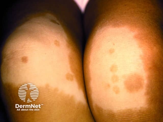
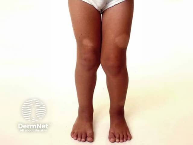
White patches of skin may also be seen on the face (particularly the chin), trunk and limbs; hands and feet are not usually affected
Often a narrow border of hyperpigmented skin surrounds the white unpigmented patches
Sometimes islands of normal or hyperpigmented skin occur within the white patches
The mild form may present with only small patches of leukoderma, whereas in more severe forms have the white forelock and larger white patches over the trunk and limbs.
Patients who are self-conscious about their appearance may benefit from the following treatments:
Dermabrasion of areas of depigmentation followed by the application of melanocyte-enriched cell suspensions
Melanocyte transplant by shaving off the top layer of skin (epidermis) and replacing it by a shave of skin from a pigmented site
Suction epidermal grafting or full-thickness punch grafts
A combination of these methods may be required and can be augmented by the addition of UV light therapy
Cosmetic camouflage techniques can cover the pigment changes of hair and skin.
What is the outcome for piebaldism?
Piebaldism is present at birth and the pigment changes usually remain unchanged throughout life. Pigmented dots and macules may develop at the margins or within the patches of leukoderma. The leukoderma may be progressive in rare cases.
Bibliography
Akarsu S, İlknur T, Avcı C, Fetil E. Piebaldism associated with café-au-lait macules and intertriginous freckling: a case report and review of the literature. Ann Dermatol. 2019;31(5):567–70. doi:10.5021/ad.2019.31.5.567. Journal
Hamadah I, Chisti M, Haider M, et al. A novel KIT mutation in a family with expanded syndrome of piebaldism. JAAD Case Rep. 2019;5(7):627–31. doi:10.1016/j.jdcr.2019.01.021. PubMed Central
Oiso N, Fukai K, Kawada A, Suzuki T. Piebaldism. J Dermatol. 2013;40(5):330–5. doi:10.1111/j.1346-8138.2012.01583.x. Journal
Richards KA, Fukai K, Oiso N, Paller AS. A novel KIT mutation results in piebaldism with progressive depigmentation. J Am Acad Dermatol. 2001;44(2):288–92. doi:10.1067/mjd.2001.112221. PubMed